
Figures & data
Figure 1 Scanning electron microscopy demonstrating mature fungal biofilms that were formed in 24-well plates. White arrows depict extracellular matrix covering and connecting the hyphae. (A) Trichophyton rubrum ATCC 28189. (B) Trichophyton mentagrophytes ATCC 11481. Reprinted from J Am Acad Dermatol, 1;80(4), Lipner SR, Scher RK, Onychomycosis: Clinical overview and diagnosis, 835–851, Copyright (2019), with permission from Elsevier.Citation4
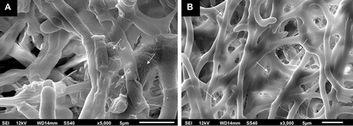
Figure 2 Patient with laboratory confirmed onychomycosis. (A) Clinical appearance of toenails with onycholysis, nail plate thickening and subungual debris. (B) Dermoscopy showing ruin-like appearance and streaks of various colors.
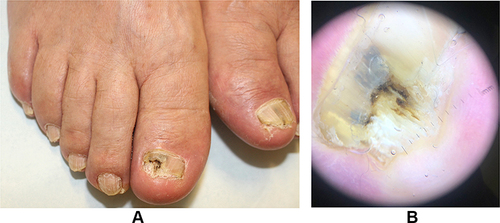
Table 1 Summary of the Diagnostic Testing Methods
Figure 3 A nail clipper is used to clip the most proximal area of onycholysis.
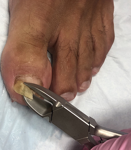
Table 2 Summary of Commonly Used Oral Onychomycosis Medications
Table 3 Summary of FDA-Approved Topical Onychomycosis Medications