Figures & data
Figure 1 Clinical manifestations of the patient. (A) Two symmetrical cicatricial alopecia patches on the scalp; (B) Recurrent follicular papules, pustules, and hemorrhagic crusts in the early stage (photographed by the patient herself); (C) Perifollicular erythema, keratosis, scales, and follicular tufts in the late stage.
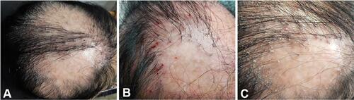
Figure 2 Histopathological findings in a vertical section showing (A) hyperkeratosis and mild epidermal hyperplasia (HE×40), (B) epithelial basal layer destruction of the hair follicle with surrounding infiltration of dense lymphocytes and histiocytes (HE×200); (C) significant plasma cell infiltration (HE×400); Horizontal section showing (D) partial destruction of hair follicles (HE×40); (E) only the structure of the arrector pill remains (HE×200).
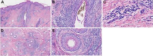
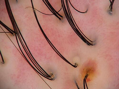
Figure 3 Trichoscopy showing follicular tufts, pustules and dilated blood vessels in an area of scarring alopecia.