
Figures & data
Figure 1 Skin biopsy from striae stained with Haematoxylin and Eosin (scale 50 μm). (A) Before treatment, (B) after 3 sessions of combined treatment, (C) after 3 sessions of fractional laser/RF, (D) after 3 sessions of PRP. Note the increase in number of fibroblasts and vascularity in all biopsies after treatment to variable degrees. Black arrows refer to fibroblasts and vascularity increasing after treatment.
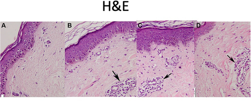
Figure 2 Skin biopsy from striae stained with Trichrome stain (scale 50 μm). (A) Before treatment, (B) after 3 sessions of combined treatment, (C) after 3 sessions of fractional laser/RF, (D) after 3 sessions of PRP. Note the increase in collagen fibres. Black arrows show increase in fibroblasts with treatment.
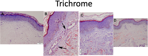
Figure 3 Skin biopsy from striae stained with Orcein (scale 50 μm). (A) Before treatment, (B) after 3 sessions of combined treatment, (C) after 3 sessions of fractional laser/RF, (D) after 3 sessions of PRP. Note the lack of increase in elastin. Black arrows point to elastic fibers.
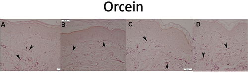
Figure 4 (A) Striae on the back before treatment, (B) improvement of striae after 3 sessions of combined treatment.
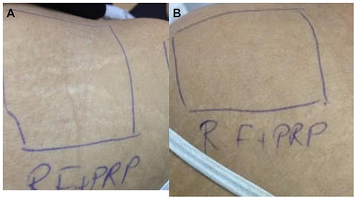
Figure 5 (A) Striae on the back before treatment, (B) improvement of striae after 3 sessions of PRP.
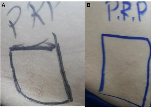
Figure 6 (A) Striae on the back before treatment, (B) improvement of striae after 3 sessions of fractional laser/RF.
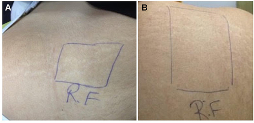
Table 1 Comparison Between Before and After in PRP
Table 2 Comparison Between Before and After in RF/CO2
Table 3 Comparison Between Before and After in Combined RF/CO2 and PRP