Figures & data
Figure 1 (A) On admission, urticarial erythema and papules on the trunk and extremities. Isolated tense blisters and bullae on the abdomen and bilateral thighs (B and C) erythema on the back and lower limbs progressed, with more new vesicles continuing to appear on the legs. (D) Erythema transformed into hyperpigmentation.
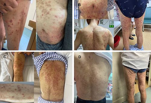
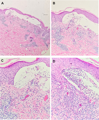
Figure 2 Pathology of skin biopsy from the lesions on the left thigh showed subepidermal blister formation with eosinophilic infiltrate, dermal vascular proliferation with lymphocytes, and eosinophil infiltration. (A) Magnification ×40, (B) magnification × 100, (C) magnification × 200, (D) magnification × 200.