Figures & data
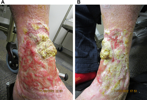
Figure 1 Ulcerative PG on the (A) anterolateral and (B) anteromedial aspects of the left leg on initial presentation, with extensive cribriform scarring and irregular overhanging borders.
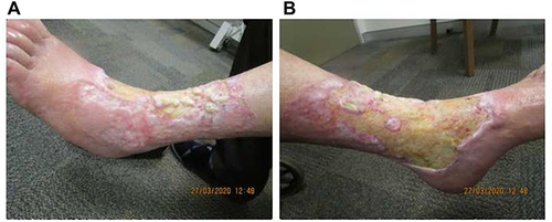
Figure 2 Re-epithelialization and reduction in ulcer size at week 8 of tildrakizumab treatment on the (A) medial and (B) anterolateral aspects of the left leg.
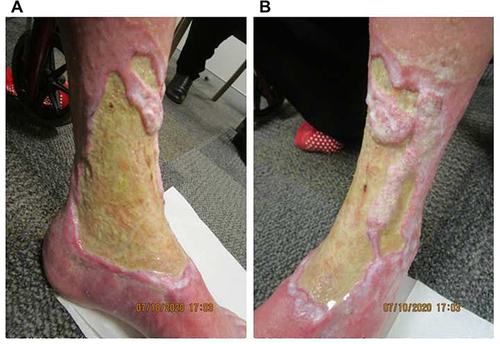
Figure 3 Excessive fibrin and thickening of the ulcer floor at week 18 of tildrakizumab treatment on the (A) medial and (B) anterolateral aspects of the left leg.
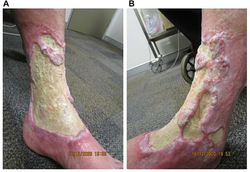
Figure 4 The underlying dermis at week 28 of tildrakizumab treatment, following judicious debridement on the (A) anteromedial and (B) anterolateral aspects of the left leg. The extensor digitorum longus tendon, previously obscured by thick fibrin, is exposed, flanked by prominent dermal proliferation.
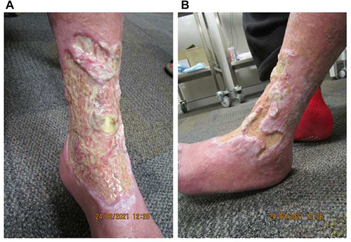
Figure 5 Re-epithelialization with bridging of advancing edges at week 55 of tildrakizumab treatment on the (A) anteromedial and (B) anterolateral aspects of the left leg.
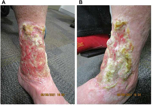
Figure 6 Further improvement at week 82 of tildrakizumab treatment on the (A) anteromedial and (B) anterolateral aspects of the left leg. Although focal areas of impetiginization are prominent, there is obvious significant further re-epithelialization.