Figures & data
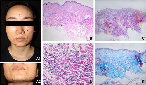
Figure 1 Multiple, skin-colored papules on face (The red arrows mark several of these typical papules). Near the left corner of the mouth, there are 2 red follicular papules (diagnosed as acne) (A1 and A2). Normal epidermis with slight fibrosis in the dermis (B) (H&E, 40×). Patchy loss of elastic fibers throughout the dermis with no correlation to hair follicles (C) (Verhoeff-Van Gieson, 40×). Prominent elastic fragmentation (D) (Verhoeff-Van Gieson, 400×). Focal hyperplasia of collagen bundles in the mid-dermis (E) (Masson staining, 40×).