Figures & data
Figure 1 (A and B) The patient had bilateral scrotal enlargement, scrotal skin thickened, significantly deepened scrotal fold, the scrotal surface can be seen needle tip to the size of a grain of rice white blisters, blisters thick wall, part of the blisters fused.
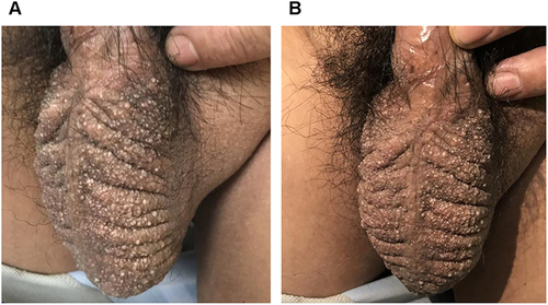
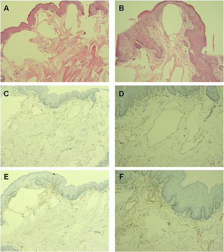
Figure 2 Skin histopathology and immunopathology examination. (A and B) Skin histopathology showed that the epidermal cells were generally normal. Several cystic spaces were composed of monolayer endothelial cells in the dermis, mostly filled with light red serous fluid. A few inflammatory cells infiltrated around the cystic spaces (HE, 100×). (C and D) Immunopathology showed CD31 positive (HE, 100×). (E and F) Immunopathology showed D2-40 positive (HE, 100×).