
Figures & data
Figure 1 Flap design and flowchart of Case 5. (A) Preoperative divided nevus position. (B) Lesion site excision extension. (C) Designed flap. (D) Immediate outcome after surgery. (E) The penis and glans position first day after surgery. (F) The penis and glans position about 6 months after surgery. (G) The penis and glans position about 27 months after surgery. (H and I), (H) Glans position, (I) Inner prepuce plate position. For the histopathology, HE staining indicated intradermal nevus under a magnification of 100×.
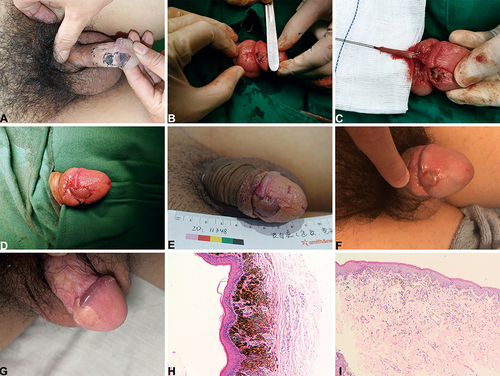
Figure 2 Flap design and flowchart of Case 8. (A) Preoperative divided nevus position. (B) Lesion site excision extension. (C) Immediate outcome after surgery. (D) The penis and glans position first day after surgery. (E) The penis and glans position 3rd day after surgery. (F) The penis and glans position about 1 month after surgery (Because of local tissue edema, the suture removal time was prolonged). (G) The penis and glans position about 6 months after surgery. (H and I), (H) Glans position, (I) Inner prepuce plate position. For the histopathology, HE staining indicated compound nevus under a magnification of 100×. Nevus cell nests at the dermo-epidermal junction and lower dermis.
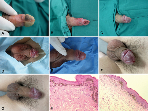
Table 1 Patient Distributions in Different Types and Severity Categories of Psychological Traits Before Operation
Table 2 The Sexual Function Scale (BMSFI and IIEF-5) Pre-Operation (Pre-) and Post-Operation (Post-) of 8 Patients
Table 3 Patient Distributions in Different Types and Severity Categories of Psychological Traits After Operation
Data Availability Statement
Data sharing not applicable to this article as no datasets were generated or analyzed during the current study.