Figures & data
Figure 1 Scattered patchy erythema and pustules on the palms of both hands and the soles (A). The palmoplantar pustules were alleviated after treatment (B).
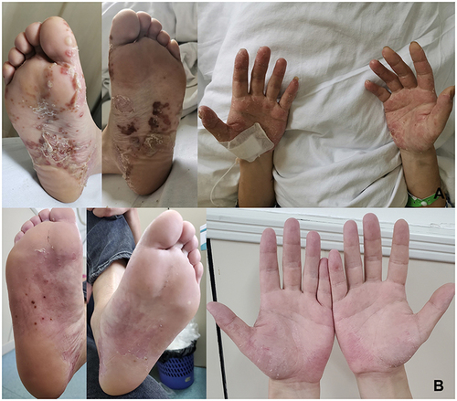
Figure 2 Skin biopsy pathology of the left hand revealed that the patient suffered from skin hyperkeratosis, with a few focal purulent exudates, epidermal hyperplasia, hypertrophy of the spinous layer and hyperplasia of the epithelium of the epidermis with lymphocytic infiltration.
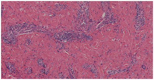
Figure 3 MRI of the thoracic spine shows abnormal signal lesions in the 5th, 8th, 9th, and 10th thoracic spine (T5, T8, T9, and T10).
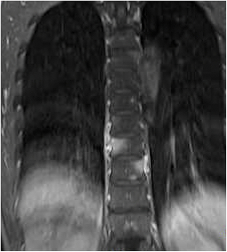
Figure 4 PET-CT presented abnormal glucose metabolism with bone alteration in the 1st right sternocostal joint, sternal body, the T5, T8, T9, and T10, and the 4th lumbar vertebra (L4), with a high possibility of a malignant lesion.
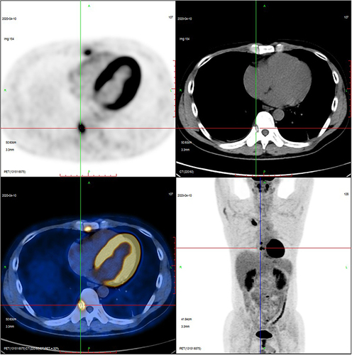
Figure 5 ECT showed that radiologically distributed thickening appeared in the 1st anterior right rib, right sternoclavicular joint, and right upper tibia ().
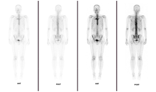
Figure 6 The patient’s vertebral biopsy pathology report did not suggest tumor cells, but only a few broken bones, sequestrums, and bone marrow.
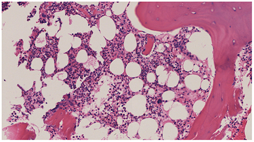
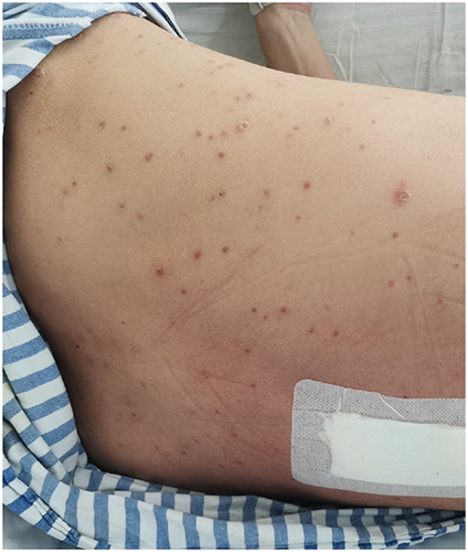
Figure 7 Pustular lesion on the anterior chest and back.