Figures & data
Table 1 Patient Characteristics
Table 2 Results: Comparison of VSS (Mean±std)
Figure 1 Comparison of VSS (A), OSAS (B) and PSAS (C). (A) comparison of VSS among the three groups before and after treatment, (B) comparison of OSAS among the three groups before and after treatment, (C) comparison of PSAS among the three groups before and after treatment. After 4 months, the improvement of VSS, OSAS and PSAS in group A and C were significantly increased compared with group B. There was no statistical difference in group A and C. *P<0.05, **P<0.01.
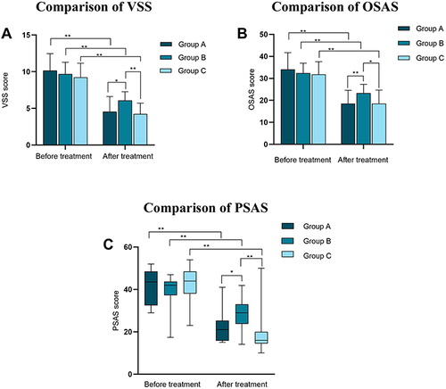
Table 3 Results: Comparison of OSAS (Mean±std)
Table 4 Results: Comparison of PSAS [Median (Interquartile Range)]
Table 5 Results: Comparison of Recurrence and Adverse Side Effects
Figure 2 Adverse side effects resulting from treatments. The most common side effect in group A, B and C was scab, scab and hyperpigmentation, respectively.
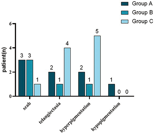
Table 6 Results: Comparison of Pain [Median (Interquartile Range)]
Table 7 Results: Comparison of Pruritus [Median (Interquartile Range)]
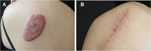
Figure 3 Example of keloid in epigastric region treated with excision followed by intralesional 5-FU and betamethasone. (A) Before treatment, (B) at 6 months of follow-up.
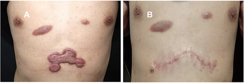
Figure 4 Example of keloid in pubic region treated with excision followed by intralesional 5-FU and betamethasone. (A) Before treatment, (B) at 6 months of follow-up.
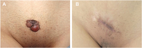
Figure 5 Example of keloid treated with intralesional 5-FU and betamethasone. (A) Before treatment, (B) at 6 months of follow-up.
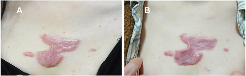
Figure 6 Example of keloid treated with excision followed by radiotherapy. (A) Before treatment, (B) at 6 months of follow-up.