Figures & data
Table 1 Demographic Data with Localization of Their Tattoo-s
Table 2 Characteristics of Tattooed Areas (Ink Colour: B = black: W = white: R = red: Y = yellow: Bl = blue: G = green: O = orange: P = pink: Br = brown: Pu = purple) and Results of Near Infra-Red Fluorescence Lymphatic Imaging-Investigation with (if Abnormal) Type of Abnormality-Ies (in-Between Brackets) (Abnl Type x)
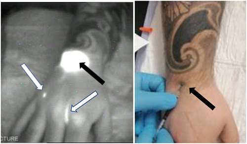
Figure 1 Normal lymphatic drainage at the level of the upper limb. On the right sided black and white figure (oblique inferior and posterior view centered on the arm), NIRFI shows fluorescent lymphatic vessel (oblique arrows) running directly under the tattooed area towards the axilla and after the intradermal injection of ICG at the level of the posterior part of the forearm under the elbow (see vertical arrows on the left sided picture) in subject j6.
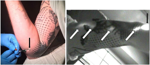
Figure 2 Normal lymphatic situation-drainage at the level of the lower limb. Anterior view on the right thigh in subject j4 showing one fluorescent lymphatic vessel running directly after the intradermal injection of ICG under the tattooed area from the injected site toward the inguinal groin.
Figure 3 Abnormality type 1. The NIRFI pictures show (left sided black and white picture) no Lymphatic Vessel (LV) (subject j9) under the tattoo at the level of the right anterior and proximal right thigh while on the opposite side (right sided black and white picture) the draining LV is seen (see black arrow) before diving deep and disappearing.
Figure 4 Abnormality type 5. The NIRFI figure (left sided) show lymphatic vessels (white arrows=subject j10) not running under the tattoo (at the level of the external and lower part of the right calf; black arrows show the ICG injected site) but seeming to bypass an obstacle and skirting the tattooed territory instead of crossing it.
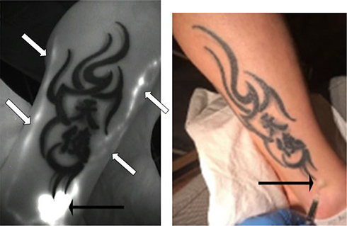
Figure 5 Abnormality type 4. The NIRFI (left sided) shows abnormal back flow from the injected site (black arrows) at the level of wrist under the tattooed area (subject p2a) toward the 2nd and 4th inter-digital spaces (white arrows).