Figures & data
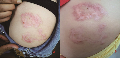
Figure 1 Clinical presentation of the patient on admission (A and B) dark red, linear, spiral patches on the lateral side of the upper arm).
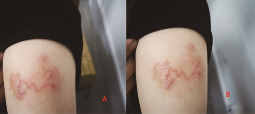
Figure 2 Dermoscopy images: (A) a tunnel with red arrows formed by larva migration. (B) high refractive dots with red arrows, which may be larval body or paws.
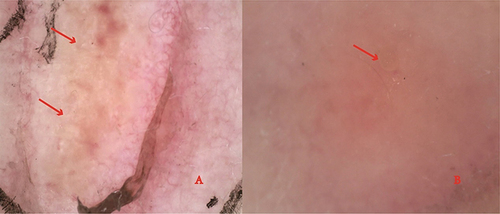
Figure 3 Reflectance confocal microscopy images: (A) tunnel in the stratum corneum or granular layer, dark oval structures corresponding to vesicles with red arrows. (B) larval body and claw-like structures with red arrows.
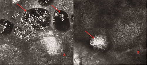
Figure 4 Clinical presentation of the patient after one week of the treatment (A and B) the rash still spread to the proximal part of the left upper arm and left shoulder with red arrows).
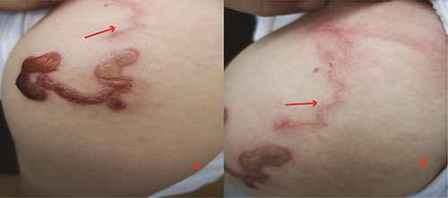
Figure 5 Clinical presentation of the patient after the treatment (A) after one month of the treatment. (B) after two months of the treatment).