
Figures & data
Figure 1 Photograph of the patient’s face before treatment. Facial swelling on the entire face and a protuberance under the inner corner of the left eye are observed. The symptoms developed on areas that received the mesotherapy injections.
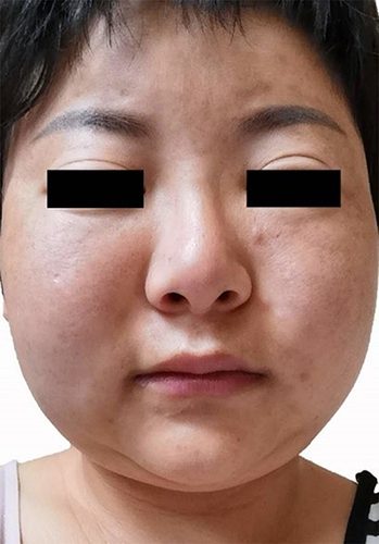
Figure 2 Echo-color Doppler (ECD) images of the patient 4 months after the mesotherapy. (a) ECD images of the left side of the face. Inhomogeneous hypoechoic nodules with distinct borders and irregular shapes were detected in the subcutaneous layer (the biggest was 5*7*6mm), with increased vascularity and enhanced echogenicity of the fat layer. (b and c) An ECD image of the right side of the face. The echogenic enhancement and increased blood flow signal were detected in the fat layer, with fewer hypoechoic nodules compared to the left side. (d) An ECD image of the forehead. Several hypoechoic nodules with distinct borders and irregular shapes were detected in the subcutaneous layer. The echogenicity of the peripheral fat layer was enhanced.
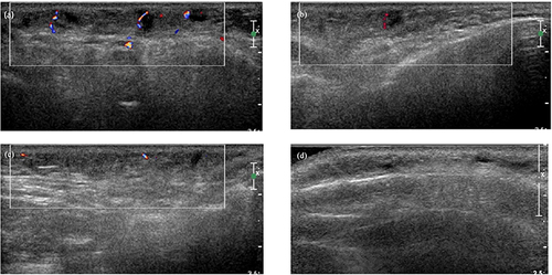
Figure 3 Photograph of the patient after 17 days of treatment in hospital with minocycline (50mg/night), 0.03% tacrolimus cream, CG (40mL /d infusion), vitamin C (10mL/d infusion) and local microwave physiotherapy.
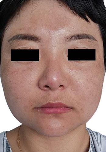
Figure 4 Photograph of the patient after 4 months of treatment at home with minocycline (100mg/ night) and 0.03% tacrolimus cream.
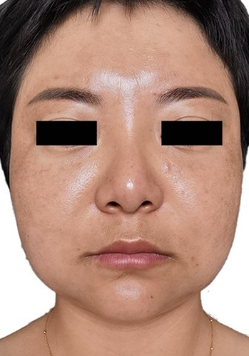
Table 1 Differential Diagnosis of Foreign Body Granuloma