Figures & data
Figure 1 Dense distribution of round and oval edematous erythema is observed on the (A) neck, front chest, and (B) back. (C and D) Portions of the erythema show blisters with the size of mung bean to soybean. Moreover, the blisters have tense walls and clear fluid and are easy to break.
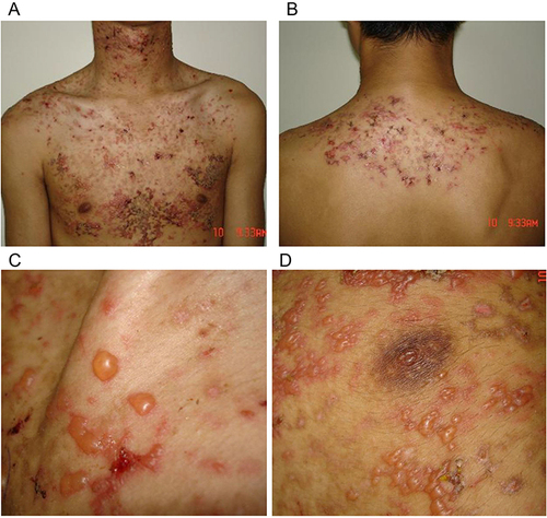
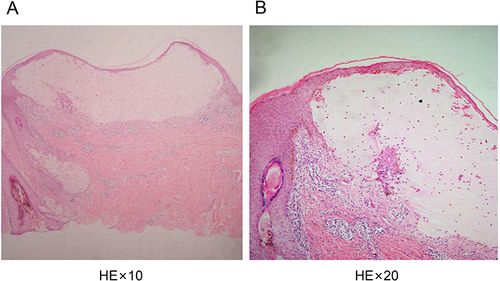
Figure 2 Skin lesions are detected as intraepidermal blisters. (A and B) The top of the blisters is composed of incomplete epidermis with scattered epidermal cells with poor keratosis. A large amount of serous fluid and low lymphocyte, neutrophil, and eosinophil levels are noted in the blisters. Additionally, a significant intracellular and intercellular edema in the lateral wall of the blisters is observed. Lastly, moderate infiltration of mixed inflammatory cells below the vesicle is observed.