Figures & data
Table 1 Mixture Components Values Used in D-Optimal Mixture Design (DMD)
Table 2 Liquid Chromatography–Mass Spectrometry (LC-MS) Identification of Compounds in Tinospora smilacina Leaf Water Extract
Figure 1 Liquid chromatography–mass spectrometry (LC-MS) chromatogram of Tinospora smilacina leaf water extract.
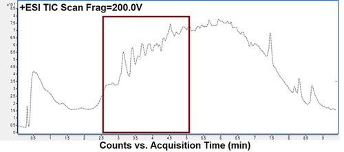
Table 3 Mixture Components and Process Factor Values Used in D-Optimal Mixture Design (DMD)
Figure 2 Three-dimensional response surface plot representing the effect of (A) Calophyllum inophyllum seed oil (CSO) (%), (B) Tween 80 (%) and (C) Tinospora smilacina water extract: high pure water (TSWE: HPW) (%) on (a) droplet size (nm) and (b) polydispersity index.
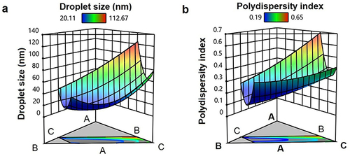
Figure 3 (a) Illustration of Tinospora smilacina water extract and Calophyllum inophyllum seed oil nanoemulsion (CTNE), (b and c) transmission electron microscopy of droplets in the CTNE. Scale bar (b) 500 nm and (c) 100 nm.
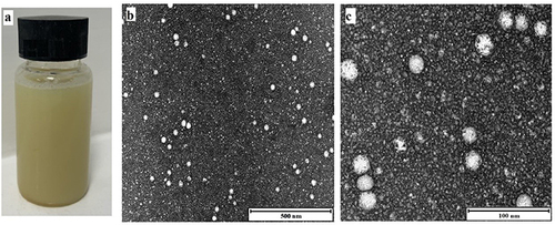
Table 4 Stability of Tinospora smilacina Water Extract and Calophyllum inophyllum Seed Oil NE (CTNE), After Four Weeks of Storage at Different Temperature
Figure 4 Cell Viability profile by MTT assay of Calophyllum inophyllum seed oil (CSO), Tinospora smilacina water extract (TSWE) and CSO nanoemulsion (CTNE) and TSWE at 24 h on hamster kidney (BSR) cell line.The x-axis indicates the NE concentration matched with the equivalent TSWE and CSO concentration content available in NE. The untreated control contained cells with media only. Each value is the mean of three replicates with standard deviation (±2 SD) and all conditions were significantly different to the untreated control analysed by two-way-ANOVA (n = 3; p < 0.05).
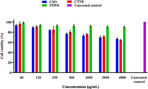
Figure 5 DPPH radical scavenging of Calophyllum inophyllum seed oil (CSO), Tinospora smilacina water extract (TSWE) and CSO nanoemulsion (CTNE) and TSWE. The DPPH results are the mean of three independent experiments with error bars ±2SD. The x-axis indicates the NE concentration matched with the equivalent TSWE and CSO concentrations available in NE.
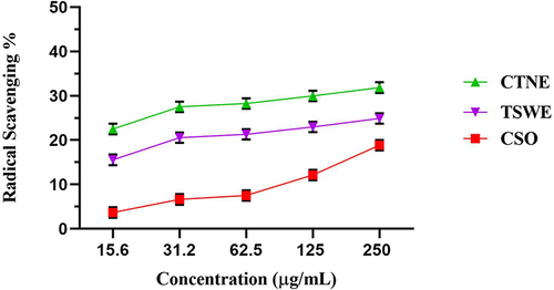
Figure 6 (a) Percentage wound closure observed for Calophyllum inophyllum seed oil (CSO), Tinospora smilacina water extract (TSWE) and CSO nanoemulsion (CTNE) and TSWE at 24 h and 48 h in hamster kidney (BSR) cells. Blank indicates untreated cells (negative control), FGF indicates Fibroblast Growth Factor (positive control) and cells with media only negative control. Each value is the mean of three individual experiments with standard deviation (±2 SD) and analysed by two-way analysis of variance (ANOVA) (n = 3; p < 0.0001). (b) Relative concentration of components available in CTNE.
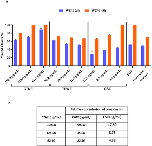
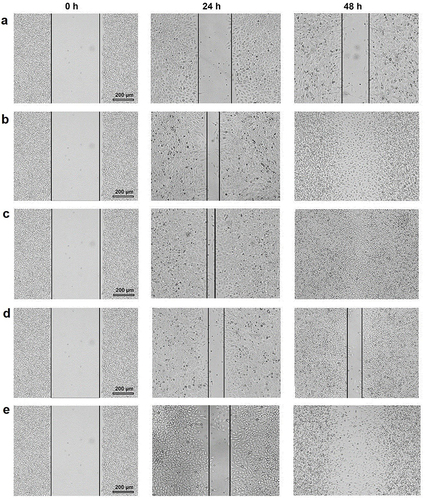
Figure 7 Wound closure photomicrograph of Calophyllum inophyllum seed oil (CSO), Tinospora smilacina water extract (TSWE) and CSO nanoemulsion (CTNE) and TSWE at 24 h and 48 h in hamster kidney (BSR) cells. (a) Untreated control. (b) Fibroblast growth factors (FGF; positive control). (c) CTNE:62.5 µg/mL. (d) TSWE:22.5 µg/mL. (e) CSO: 4.3 µg/mL. Black solid lines represent the wound size (μm) of the BSR cell line monolayer.