Figures & data
Table 1 The Positive Rate and Intensity of CD2AP Expression in 25 Cases of Chronic Eczema and 18 Cases of Normal Skin Around Eczema
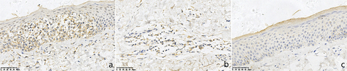
Figure 1 Expressions of CD2AP in chronic eczema lesions and the perilesional normal skin at the edge (SP *400). (a) Epidermic of skin lesion. (b) Dermis of skin lesion. (c) Perilesional normal skin. (a) More CD2AP+ monocyte-like cells with brown cytoplasm were observed in the epidermal of chronic eczema lesions. (b) More CD2AP+ monocyte-like cells with brown cytoplasm were observed in the dermal papillary layer of chronic eczema lesion. (c) Almost no of CD2AP+ monocyte-like cells with brown cytoplasm were observed in both epidermal and papillary layer of perilesional normal skin of the lesion.
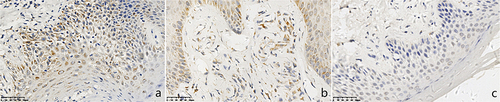
Table 2 The Positive Rate and Intensity of CD123 Expression in 22 Cases of Eczema and 18 Cases of Normal Skin Around Eczema
Figure 2 Expressions of CD123 in chronic eczema lesions and the perilesional normal skin at the edge (SP *400). (a) Epidermic of skin lesion. (b) Dermis of skin lesion. (c) Perilesional normal skin. (a) A few CD123+ monocyte-like cells with brown cytoplasm were observed in the epidermal of chronic eczema lesions. (b) More CD123+ monocyte-like cells with brown cytoplasm were observed in the dermal papillary layer of chronic eczema lesion. (c) Almost no of CD123+ monocyte-like cells with brown cytoplasm were observed in both epidermal and papillary layer of perilesional normal skin of the lesion.
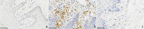
Table 3 The Positive Rate and Intensity of TLR7 Expression in 17 Cases of Chronic Eczema and 13 Cases of Normal Skin Around Eczema
Figure 3 Expressions of TLR7 in chronic eczema lesions and the perilesional normal skin at the edge (SP *400). (a) Epidermic of skin lesion. (b) Dermis of skin lesion. (c) Perilesional normal skin. (a) A few TLR7+ cells with brown cytoplasm were observed in the epidermal of chronic eczema lesions. (b) A few TLR7+ cells with brown cytoplasm were observed in the dermal papillary layer of chronic eczema lesion. (c) Few TLR7+ cells with brown cytoplasm were observed in both epidermal and papillary layer of perilesional normal skin of the lesion.
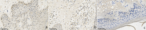
Table 4 The Positive Rate and Intensity of TLR9 Expression in 16 Cases of Chronic Eczema and 14 Cases of Normal Skin Around Eczema
Figure 4 Expressions of TLR9 in chronic eczema lesions and the perilesional normal skin at the edge (SP *400). (a) Epidermic of skin lesion. (b) Dermis of skin lesion. (c) Perilesional normal skin. (a) A few TLR9+ cells with brown cytoplasm were observed in the epidermal of chronic eczema lesions. (b) A few TLR9+ cells with brown cytoplasm were observed in the dermal papillary layer of chronic eczema lesion. (c) Few TLR9+ cells with brown cytoplasm were observed in both epidermal and papillary layer of perilesional normal skin of the lesion.
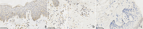
Table 5 The Positive Rate and Intensity of IRAK1 Expression in 23 Cases of Chronic Eczema and 20 Cases of Normal Skin Around Eczema
Figure 5 Expressions of IRAK1 in chronic eczema lesions and the perilesional normal skin at the edge (SP *400). (a) Epidermic of skin lesion. (b) Dermis of skin lesion. (c) Perilesional normal skin. (a) More IRAK1+ cells with brown cytoplasm were observed in the epidermal of chronic eczema lesions. (b) More IRAK1+ cells with brown cytoplasm were observed in the dermal papillary layer of chronic eczema lesion. (c) Almost no of IRAK1+ cells with brown cytoplasm were observed in both epidermal and papillary layer of perilesional normal skin of the lesion.
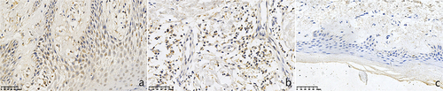
Table 6 The Positive Rate and Intensity of IRF7 Expression in 22 Cases of Chronic Eczema and 16 Cases of Normal Skin Around Eczema
Figure 6 Expressions of IRF7 in chronic eczema lesions and the perilesional normal skin at the edge (SP *400). (a) Epidermic of skin lesion. (b) Dermis of skin lesion (c) Perilesional normal skin. (a) More IRF7+ cells with brown cytoplasm were observed in the epidermal of chronic eczema lesions. (b) More IRF7+ cells with brown cytoplasm were observed in the dermal papillary layer of chronic eczema lesion. (c) Almost no of IRF7+ cells with brown cytoplasm were observed in both epidermal and papillary layer of perilesional normal skin of the lesion.