Figures & data
Figure 1 Clinical presentations at admission. (A and B) Hypertrophic dark red plaques were seen in the head-face-neck area and torso, which had partially merged together.
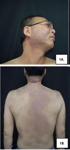
Figure 2 (A) (×100) and (B) (×200): Obvious perivascular infiltration of clumps of lymphocytes, plasma cells, and eosinophils was observed from the superficial to deep dermis; (C) (×100), (D) (×100), (E) (×100) and (F) (×100): IHC examinations of CD138, Ki-67, IgG, and IgG4 respectively, with IgG4/IgG exceeding 40%.
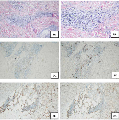
Figure 3 (A) Multiple lymph nodes and several enlarged ones were found near the bilateral neck (IIA), bilateral armpits, and iliac vessels of bilateral pelvic wall, with enhanced FDG metabolism; (B) Soft tissue density occupancy in the intermuscular spaces of left retrobulbar muscle cone was observed together with improved FDG metabolism, which was consistent with inflammatory pseudotumor changes within the eye frame in combination with his medical history.
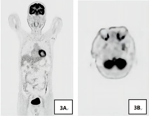
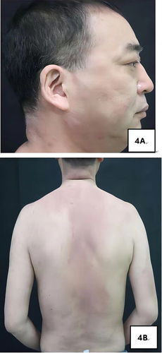
Figure 4 Lesion manifestations after 3 months of treatment. (A and B) After 6 days of treatment, the skin lesions improved and partially resolved, with mild lesions remaining and post-inflammatory hyperpigmentation.