Figures & data
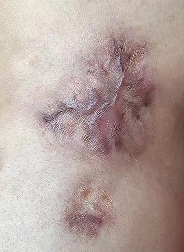
Figure 1 Clinical photograph of the patient: Initial photograph of the patient.
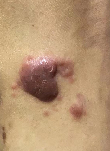
Figure 2 Clinical photograph of the patient: after topical injections of compound betamethasone injections 1 mL x 4 times.
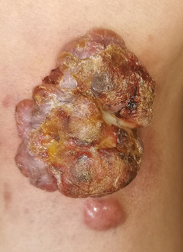
Figure 3 Histopathology of skin lesions: Diffuse infiltration of tumour cells in the dermis and subcutaneous tissues, with large tumour cells, abundant cytoplasm, round or irregular nuclei, obvious nucleoli, obvious heterotypes, obvious interstitial phenomenon, and focal infiltration of surrounding lymphocytes (HEx100).
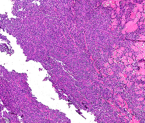
Figure 4 Histopathology of skin lesions: with large tumour cells, abundant cytoplasm, round or irregular nuclei, obvious nucleoli, obvious heterotypes, obvious interstitial phenomenon (HEx200).
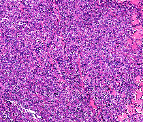
Figure 5 Mmunohistochemistry: CD30 (+++)(IHC x100).
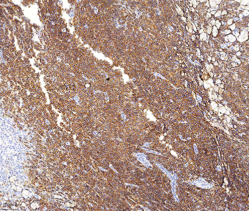
Figure 6 Mmunohistochemistry: CD30 (+++) (IHC x200).
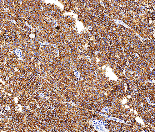
Figure 7 Clinical photograph of the patient: After 6 rounds of chemotherapy with the CHOP regimen.