Figures & data
Figure 1 Erythemas, lichenoid papules, scaly patches on his face, upper extremities and trunk (a and b). The scattered skin tags were mainly located on his upper extremities and trunk (b–d).
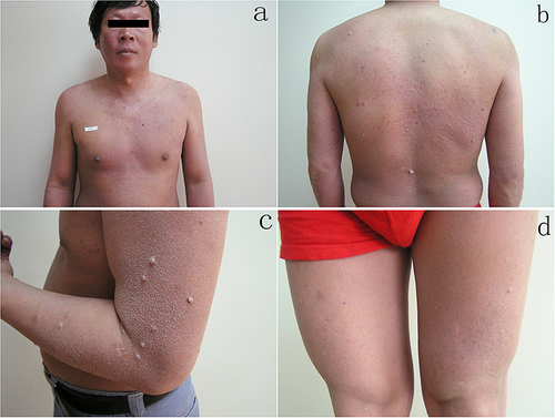
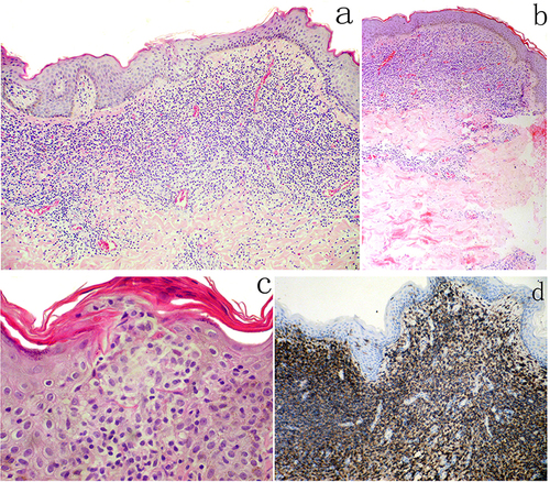
Figure 2 The biopsies from the papule (a) and the skin tag (b) show a dense, band-like lymphocytic infiltrate in the papillary and mid-reticular dermis. The super-basal epidermis contained both single and small collections of lymphoid cells (Pautrier’s collections) (c). Most of lymphoid cells are CD45RO-positive cells (d).