Figures & data
Figure 1 (A and B) Poorly demarcated greyish-brown confluent macules and patches associated with fine rough scales on the right cheek and the left cheek respectively. Lesions on the right side are darker and more diffuse compared to the left side. (C and D) Sites of the lesions after using ivermectin 1% cream daily at 3-month follow-up, significant improvement in discoloration and texture on the right cheek and the left cheek respectively.
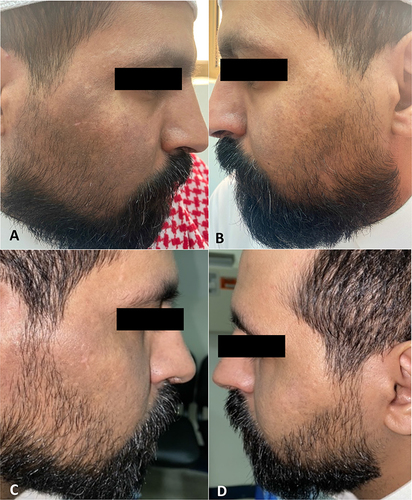
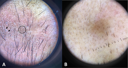
Figure 2 (A) dermoscopic picture showing white gelatinous filaments protruding through follicular openings “demodex tails” (arrows), and reticular perifollicular pigmented network (circle). (B) dermoscopic picture showing improvement after 3 months of treatment with ivermectin 1% cream.