Figures & data
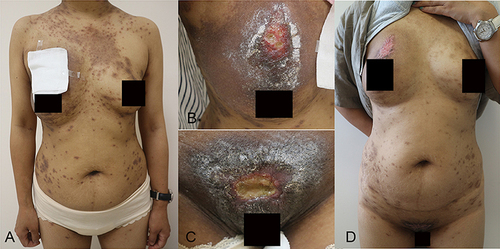
Figure 1 Clinical manifestations in a 38-year-old female with PG before treatment. Scattered ulcers were seen on the patient’s pubic area, left thigh, right buttock, right breast, and eyebrow, with red granulation tissue and yellow necrotic debris on the surface of the ulcers and edema at the elevated border. (A–E) Histopathology of a PG lesion from the right thorax (stained with haematoxylin and eosin; magnifications: (F) ×40, ((G and H) ×200). The epidermis is hypertrophic, spongy and oedematous, with neutrophil migration into the epidermis (F). The dermis is locally infiltrated with neutrophils forming areas of cerebral swelling and necrosis, with surrounding areas of granulomatous inflammation and infiltration of inflammatory cells such as lymphocytes, plasma cells and eosinophils (G), with necrosis of the vessel wall and spillage of large red blood cells (H).
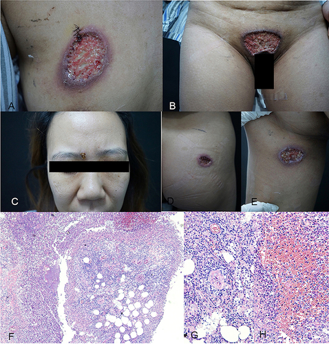
Figure 2 Clinical manifestations in a 38-year-old female with PG and eczema. Scattered ulcers with a small amount of pus on the surface were seen on the right chest and vulva. The ulcers were surrounded by erythema and hyperpigmentation. There were scattered erythema, claw marks, and crusts on the trunk. (A–C) and after 4 months of treatment with baricitinib. Pinkish scarring was seen on the right breast and vulva. There was scattered pigmentation on the trunk. (D).