Figures & data
Figure 1 Clinical photo showed an irregular and lobulated mass lesion spanning across the right eyebrow region with a small depression and hyperpigmented area in the surface (white arrow).
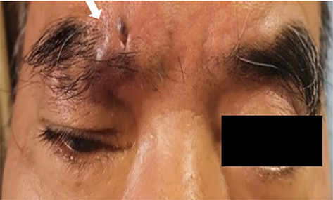
Figure 2 (A) CT axial image revealed an ill-defined and irregular soft tissue density shadow spanning across the eyebrow region (white arrow). The CT value was 40HU.The shape and size of the eyeballs, extraocular muscles and optic nerve on both sides were normal and symmetrical. No abnormality was observed in the orbital bone. (B) Sagittal reconstruction from the CT scan image showed the lesion locating above the right orbit to the forehead (white arrow). The lesion exhibited on MRI as an inhomogenous hypointense (white arrow) on T1-weighted imaging (T1WI) (C) and hyperintense (white arrow) on T2-weighted imaging (T2WI) (D) with unclear boundary. (E and F) Axial and sagittal contrast-enhanced T1WI showed a heterogeneous enhancement (white arrows).
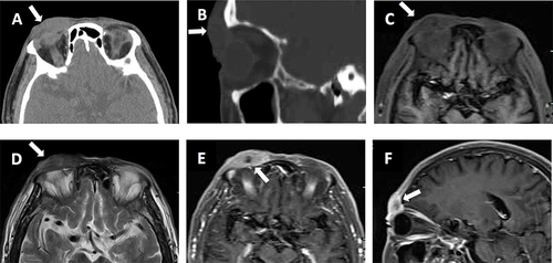
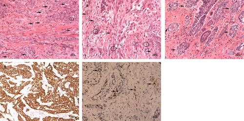
Figure 3 Postoperative histopathological examination of resected lesion showed diffuse infiltrative growth of the mass with hematoxylin and eosin (H&E) stain at 200× magnification. (A) In the typical zone of the lesion, a large number of nest-like heterotypic cells were seen within the fibrous stroma of dermis. Some cells contained large, oval or round nuclei and vacuolated clear cytoplasm (black arrows), and some had vesicular nuclei with small nucleoli could be observed and nuclear division easily visualized (black circle). (B) Spindle sarcoma-like cells were visualized in part of the sarcomatous zone of the lesion (black arrows) which had a transition between the nest-like component zone (black circles). (C) In the BCC zone of the lesion, some tumor cells in the superficial dermis were found to be small nest-like (black arrows), and some basal cells arranged in a palisading pattern like BCC. IHC staining (EnVision Method) was performed on the neoplasm and revealed CK5/6 strongly positive at 100× magnification (D) and AR focally positive (black arrows) at 200× magnification (E).