Figures & data
Figure 1 Clinical appearance of the skin lesion. (A) Scattered erythema and infiltrative plaques on the head, isolated and not fused. (B and C) Numerous papules and nodules on the patient’s trunk and limbs, with well-demarcated.
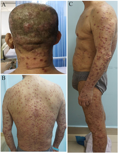
Figure 2 Histological findings of plaque on the head. (A and B) Prominent plasma cell infiltration of the dermis (HE, ×400).
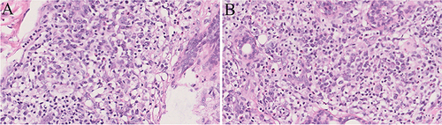
Figure 3 Immunohistochemistry results. (A–L) Infiltrating plasmacytes were CD3, CD4, CD20, CD8, CD68, CD163, CD138, Ki-67, Kappa, Lambda, IgG and IgG4 positive (×200).
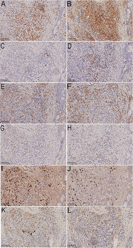
Figure 4 Clinical appearance after 6 months of treatment.
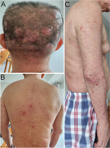
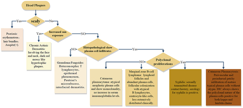
Figure 5 Algorithm for head plaques finding.