Figures & data
Figure 1 Hematoxylin and eosin-stained biopsy lesion taken from FD-diseased areas of the scalp showing follicular obliteration by acute and chronic inflammatory cell infiltrate (blue arrow) with dermal scarring (red arrows) magnification x2 (A) and inflammatory cell infiltrate composed of mature lymphocytes and neutrophils (green arrow) magnification x10 (B).
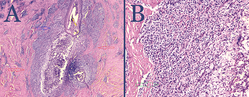
Figure 2 Patient 1: Postoperative FD plaque involving the vertex, mid-scalp extending to the frontal scalp, immediately after excision (A); tension Suture guards to enable high-tension horizontal mattress suturing with less tissue tear-through or risk of tissue strangulation. Showing side (top) and top (bottom) views.(B), and postoperative FD plaque involving the vertex, mid-scalp extending to the frontal scalp immediately after applying high-tension sutures with guards in a horizontal mattress fashion and long suture tails to enable retightening as needed. The wound bed is filled with bacitracin antibiotic ointment (C).
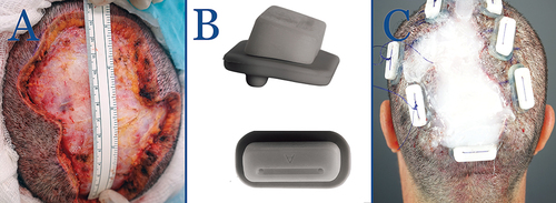
Figure 3 Patient 3: Postoperative FD plaque involving the vertex, and mid-scalp, immediately after applying high-tension sutures with guards in a horizontal mattress fashion. The wound bed is filled with bacitracin antibiotic ointment. (A) At eight weeks postoperatively, a small STSG derived from the anterior thigh is used to close the residual open wound (red arrow). Tenting of skin from tension suture effect is visible (yellow arrows) (B).
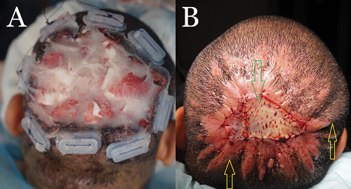
Table 1 Demographic and Pre-Surgical Data on Five Patients That Underwent Surgical Excision of FD Lesions with SIH Aided by Guarded High-Tension Suturing
Table 2 Surgical and Post-Surgical Data on Five Patients That Underwent Surgical Excision of FD Lesions with SIH Aided by Guarded High-Tension Suturing
Figure 4 Patient 1: Preoperative FD plaque involving the vertex, mid-scalp extending to the frontal scalp. (A) Thirteen months after complete excision of FD lesion and healing by second intention, aided by high-tension sutures with guards and a minor skin graft (B).
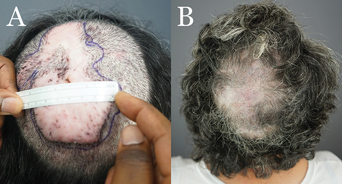
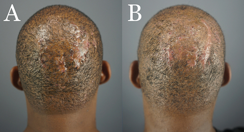
Figure 5 Patient 2: Preoperative FD plaque involving the vertex, mid-scalp extending to the frontal scalp, and acne keloidalis nuchae in the nape area (A) and nineteen months after complete excision of FD lesion and healing by second intention, aided by guarded high-tension sutures and a minor STSG (Green arrow). The tenting skin has flattened out (yellow arrows (B).
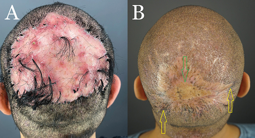
Figure 6 Patient 3: Preoperative FD plaque involving the entire vertex. (A) and eight months after complete excision of the FD lesion and healing by second-intention, aided by guarded high-tension sutures and a minor skin graft(B).
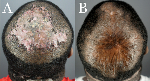
Figure 7 Patient 4 with FD plaque involving the right parietal prominence area before surgical excision and AKN lesions in the nape zone. (A) and four months after complete excision of the FD lesion and healing by second-intention, aided by guarded high-tension sutures and a minor skin graft (B).
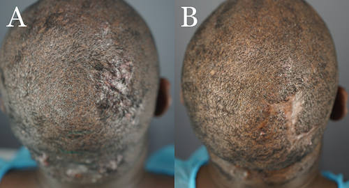
Figure 8 Patient 5 with FD plaque involving the right parietal prominence area before surgical excision. (A) and nine months after complete excision of the FD lesion and healing by second-intention, aided by guarded high-tension sutures (B).