
Figures & data
Figure 1 Skin lesion performance of abrocitinib before and after treatment of patients with EPPP. (a) Skin lesions prior to the treatment; (b) Lesion at higher magnification; (c) Significant improvement in the degree of skin lesions after one month of treatment with abrocitinib.
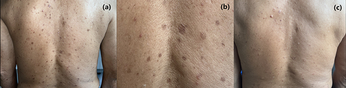
Figure 2 Pathological presentation of the patient. (HE, ×100). The epidermis is mildly hyperplastic, with several keratin-filled depressions showing a tendency to form a conical plate with vacuolated epithelial cells and a partial lack of a granular layer. Lymphocyte infiltration can be seen around dermal vessels (a and b).
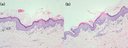
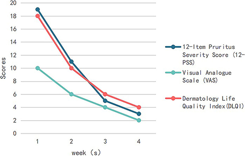
Figure 3 Questionnaire evaluation of abrocitinib before and after treating patients with EPPP. Pruritus was assessed using the visual analog scale (VAS) and the 12-item pruritus severity scale (12-PSS), and data on quality of life were assessed using the dermal life quality index (DLQI).
Data Sharing Statement
Data sharing is not applicable to this article as no datasets were generated or analyzed during the current study.