Figures & data
Table 1 Clinical Data and Treatment of EPF Patients
Figure 1 Clinical pictures: (A–I) Follicular papules with a diameter of about 1–2 mm are seen based on erythema on the face and trunk and expand eccentrically to the periphery in a circular or creeping pattern, and pustules are seen in some patients.
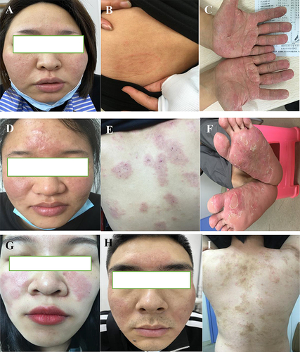
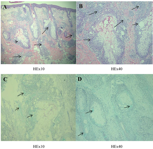
Figure 2 Histopathology: (A) Mild epidermal hyperplasia, lymphocytic and eosinophilic infiltration around the superficial middle dermal vessels and appendages (black arrow), (B) eosinophilic and neutrophilic abscesses seen in local hair follicles (black arrow). (C and D) Acidic mucin deposits around the hair follicle and the hair follicle’s sebaceous glands (black arrow).