
Figures & data
Figure 1 (A–C) Skin condition before and after treatment. Upon admission, multiple yellowish-red or reddish-brown papules and nodules almost spread throughout the body, with clear boundaries and partial fusion. The back area was the most severe (A). After the completion of 4 courses of CHOP+ lenalidomide, the rash subsided significantly, with significant pigmentation on the back (B). Two courses of chemotherapy were completed after recurrence, and the rash almost completely subsided with only a small amount of pigmentation on the back (red arrow, biopsy scar after recurrence) (C). (D) [18F] FDG-PET/CT showed considerable metabolic increases in the skin (the red arrows point to some areas exhibiting heightened metabolic activity). Areas including the upper limbs (upper arm), lower neck, body (back), and the skin/subcutaneous area of the upper thighs of both sides within the detection range, and some of them were accompanied by small nodular shadows. The possibility of malignant tumor lesions was considered. Immunohistochemical studies demonstrated that the histiocytes were positive for CD1a (E), CD68 (F) and S-100 (G) and negative for Langerin (H).
![Figure 1 (A–C) Skin condition before and after treatment. Upon admission, multiple yellowish-red or reddish-brown papules and nodules almost spread throughout the body, with clear boundaries and partial fusion. The back area was the most severe (A). After the completion of 4 courses of CHOP+ lenalidomide, the rash subsided significantly, with significant pigmentation on the back (B). Two courses of chemotherapy were completed after recurrence, and the rash almost completely subsided with only a small amount of pigmentation on the back (red arrow, biopsy scar after recurrence) (C). (D) [18F] FDG-PET/CT showed considerable metabolic increases in the skin (the red arrows point to some areas exhibiting heightened metabolic activity). Areas including the upper limbs (upper arm), lower neck, body (back), and the skin/subcutaneous area of the upper thighs of both sides within the detection range, and some of them were accompanied by small nodular shadows. The possibility of malignant tumor lesions was considered. Immunohistochemical studies demonstrated that the histiocytes were positive for CD1a (E), CD68 (F) and S-100 (G) and negative for Langerin (H).](/cms/asset/aafcd0b3-0022-4769-b7a0-ed632f1a8cc9/dcci_a_12296597_f0001_c.jpg)
Figure 2 The timeline of this case report.
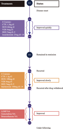
Table 1 Reported Cases of IDCT with Cutaneous Symptoms Treated with Chemotherapy Medications