Figures & data
Figure 1 Clinical manifestations of LEP in the patient. (A) Multiple cutaneous ulcer and scarring alopecia. (B) Serous crust overlying indurated erythematous plaque on the forehead. (C and D) Clinical improvement after three months of follow up.
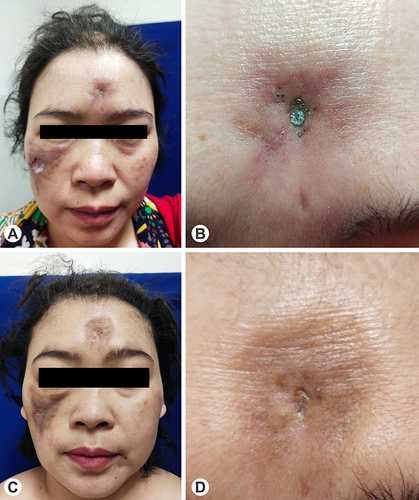
Figure 2 Cutaneous ulcers on the vertex and right cheek. (A and B) Prior to treatment. (C and D) After the application of modern wound dressing and topical antibiotic in the first month of follow up. (E and F) After the treatment with oral hydroxychloroquine in the third month of follow up.
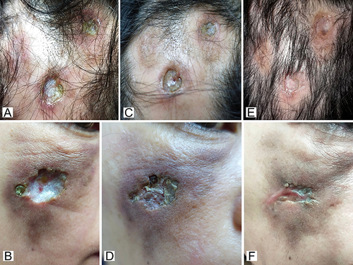
Figure 3 Histopathological findings with HE-staining revealed perivascular (red circle), periadnexal (yellow circle), and periadiposal (blue circle) lymphocytic, plasmacytic, and eosinophilic infiltration.
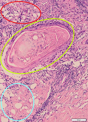
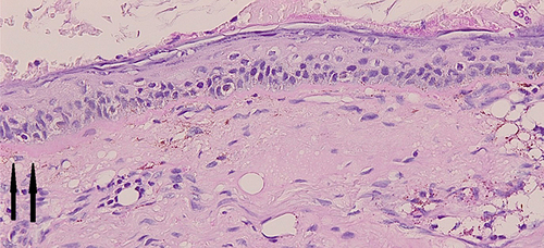
Figure 4 PAS-staining revealed thickening of the basement membrane pointed with black arrows.