Figures & data
Figure 1 Clinical images and relevant examination results obtained during the patient’s initial visit to our hospital. (a and b) Skin examination unveiled infiltrative symmetrical Orange or brown patches with distinct borders on the inner canthi as well as upper and lower eyelids. The surface was smooth, devoid of scales, atrophy, erosion, or necrosis, and exhibited no tenderness. (c) The epidermis displayed mild hyperplasia, while the dermis exhibited a conglomerate of foam cells, histiocytes, and multinucleated giant cells. Additionally, a minor lymphocytic infiltration was observed around the dermal blood vessels and adnexal structures adjacent to the conglomerate (H and E×50). (d) Intense lymphoplasmacytic infiltration was observed (H&E, magnification ×200). (e) Negative result for S100 immunohistochemistry (magnification ×200). (f) Absence of CD1a immunohistochemical staining (magnification ×200). (g) Positive immunohistochemical staining for CD68 (magnification ×200). (h and i) The axial contrast-enhanced orbital CT scan revealed no abnormalities in either eye. Swelling of the subcutaneous soft tissue was noted in the bilateral maxillofacial region and eyelids, with a subtle prominence on the right side.
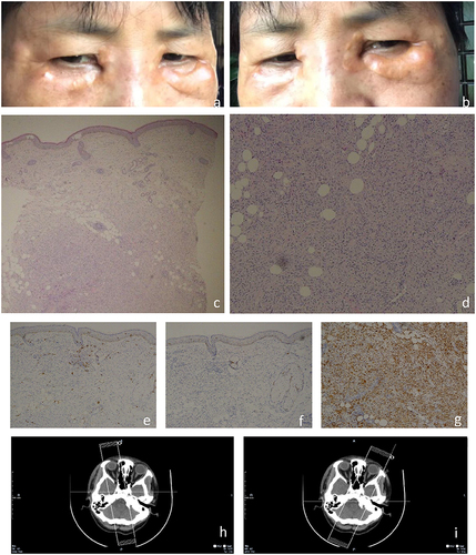
Figure 2 Clinical picture of the patient’s sister. Soft, yellow plaques were observed at the medial canthus of both the upper and lower eyelids.