
Figures & data
Figure 1 Erythema, papules, scales and excoriation on the waist.
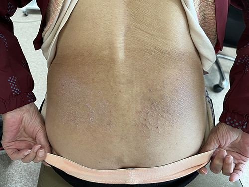
Figure 2 Psoriasis- like lesions: demarcated scaly erythematous lesions.
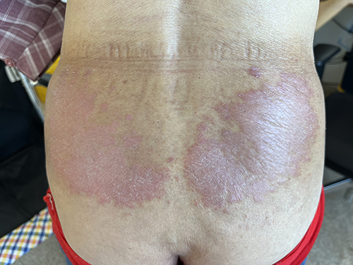
Figure 3 Histopathology showed psoriasiform acanthosis with a diminished granular layer, lymphocyte infiltration in the superficial dermis. (HE stain, magnification 4*10).
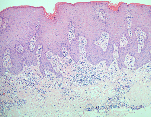
Figure 4 Topical calcipotriol and betamethasone ointment was applied to the psoriasis-like lesions.
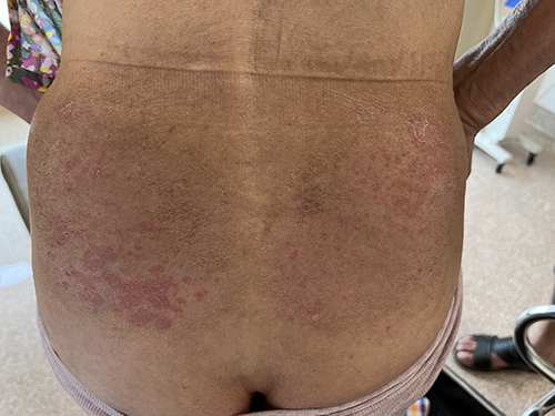
Figure 5 (A–D) Erythema, papules, scales on the shoulder (A), back (B), lower limbs (C) and popliteal fossa (D).
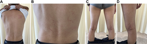
Table 1 The Relevant Reports from the Past 5 Years