75
Views
2
CrossRef citations to date
0
Altmetric
Review

Erosive pustular dermatosis (chronic atrophic dermatosis of the scalp and extremities)

, &
Pages 177-182 | Published online: 11 Jul 2013

Figures & data

Figure 1 (AC) Chronic eosinophilic dermatosis of the scalp in a 76 year-old male. (A) Overview. (B) Detail of crusted and erosive lesion with overgranulation. No signs of reepithelialization. (C) Excision and split-skin transplant. Ten days after transplantation, a stable transplant without recurrence is seen.

Figure 1 (A–C) Chronic eosinophilic dermatosis of the scalp in a 76 year-old male. (A) Overview. (B) Detail of crusted and erosive lesion with overgranulation. No signs of reepithelialization. (C) Excision and split-skin transplant. Ten days after transplantation, a stable transplant without recurrence is seen.

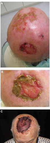
Table 1 Differential diagnosis of erosive pustular dermatitis of the scalp (EPDS)

Figure 2 A 57-year-old patient with chronic actinic damage, multiple actinic keratoses and a T2 squamous cell carcinoma on the scalp. Field cancerization is an important differential diagnosis to erosive pustular dermatosis of the scalp.

Figure 2 A 57-year-old patient with chronic actinic damage, multiple actinic keratoses and a T2 squamous cell carcinoma on the scalp. Field cancerization is an important differential diagnosis to erosive pustular dermatosis of the scalp.

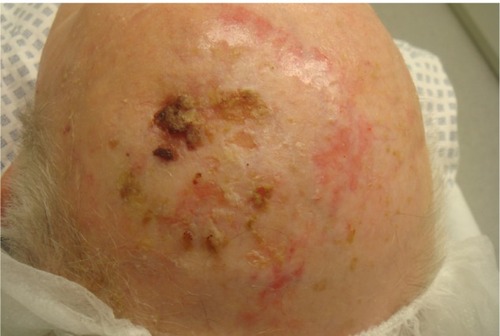
Table 2 Therapeutic opportunities in erosive pustular dermatosis of scalp and leg