Figures & data
Table 1 List of selected tissue biomarkers in melanoma
Figure 1 MiTF accurately enumerates the melanocytes in the epidermis of chronic sun exposed skin and in lesions of lentigo maligna.
Abbreviations: H&E, hematoxylin and eosin; IHC, immunohistochemistry.
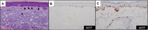
Figure 2 Evaluation of bread-loafed cross-sections of peripheral margins allows for comparison of the background increased density of melanocytes, reflective of chronic sun damage (at the periphery), with area of lentigo maligna (at the center).
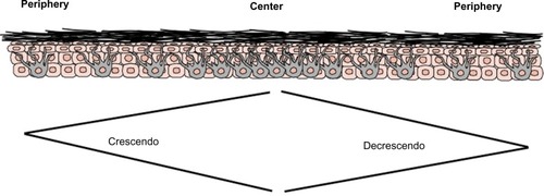
Figure 3 Sox10 identifies rare tumor cells in heavily regressed areas of melanoma.
Abbreviations: H&E, hematoxylin and eosin; IHC, immunohistochemistry.
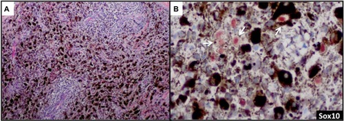
Figure 4 Detection and confirmation of lymphovascular invasion.
Abbreviations: H&E, hematoxylin and eosin; IHC, immunohistochemistry; LVI, lymphovascular invasion.
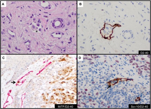
Figure 5 Detection of mitotic figures and G2+ tumor nuclei with histone markers.
Abbreviation: IHC, immunohistochemistry.
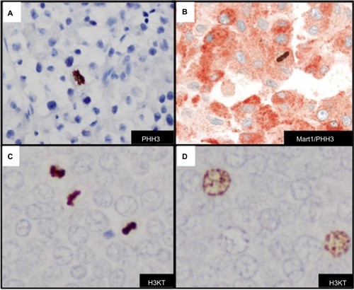
Figure 6 Immunohistochemcial evaluation of BRAFV600E mutation status in melanoma.
Abbreviations: AEC, 3-amino-9-ethylcarbazole; DAB, diaminobenzidine; H&E, hematoxylin and eosin; IHC, immunohistochemistry.
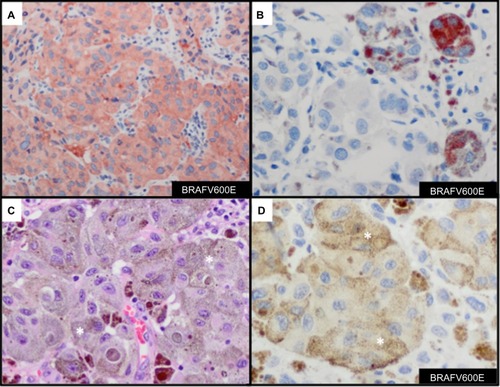
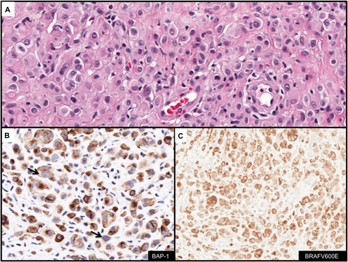
Figure 7 Immunohistochemcial evaluation of BAP-1 and BRAFV600E status in melanocytic lesions.
Abbreviations: H&E, hematoxylin and eosin; IHC, immunohistochemistry.