Figures & data
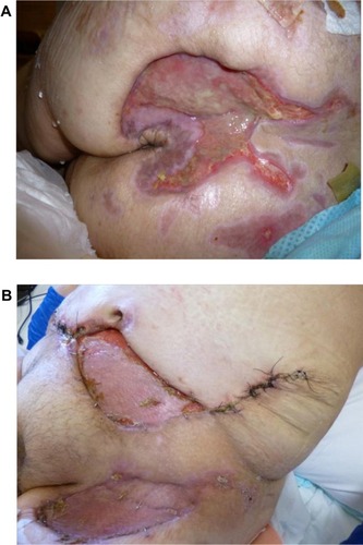
Figure 1 (A) Sacral pyoderma gangrenosum after multiple debridements. (B) Huge abdominal and left thigh wound defect with typical violaceous borders post-debridement.
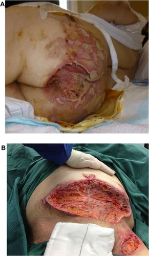
Figure 2 Histology of incisional biopsy of abdominal ulcer. (A) Extensive skin ulceration is shown (H&E stain; original magnification ×10). (B) Higher-powered view of ulcer being covered by inflamed crust and infiltrated by a heavy collection of acute inflammatory cells (neutrophils) (H&E stain; ×20).
Abbreviation: H&E, hematoxylin and eosin.
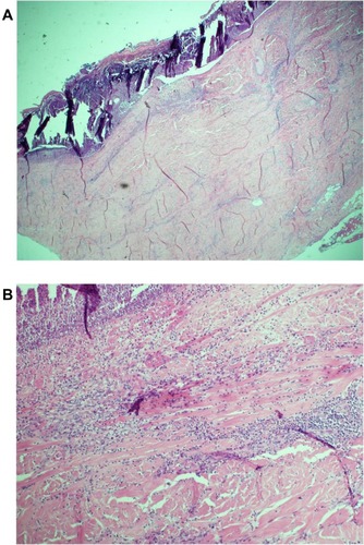
Figure 3 After treatment with immunosuppressive medications, appearance of sacral (A) and abdominal and left thigh (B) pyoderma gangrenosum.