
Figures & data
Table 1 Differential diagnosis of the unspecific ulcerative lesions of pyoderma gangrenosum
Figure 1 (A) Ulcerative stage of a pyoderma gangrenosum lesion, showing the typical violaceous, raised undermined border, with a granulation base, surrounded by a erythematous halo. (B) Healing stage, after 4 days therapy with systemic corticosteroids and cyclosporine, showing a decrease in the surrounding inflammation.
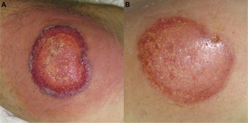
Figure 2 (A) Ulcerative stage of classical and rapidly progressive pyoderma gangrenosum ulcer, showing an elevated, violaceous, undermined border, with a necrotic and hemorrhagic base. (B) The healing stage ulcer, 3 months after systemic therapy, presenting the “Gulliver” sign; ulcer base containing granulation tissue, and necrotic tissue in a lesser extent.
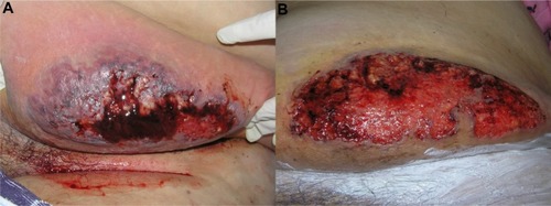
Figure 3 Characteristic cribriform scar after healing of pyoderma gangrenosum.
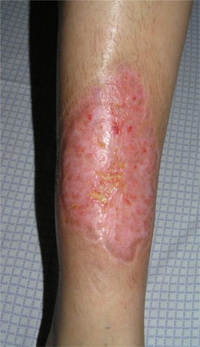
Figure 4 Bullous variant of pyoderma gangrenosum, in a patient with acute myeloid leukemia, presenting overlap features with bullous variant of Sweet’s syndrome.
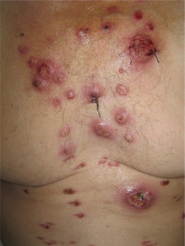
Table 2 Proposed diagnostic criteria for classic ulcerative pyoderma gangrenosum, according to Su et al,Citation4 diagnosis would require two major and at least two minor criteria