Figures & data
Figure 1 Potassium hydroxide 10% mount.
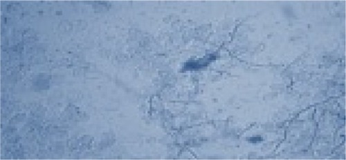
Figure 2 Trichophyton violaceum (violet pigment).
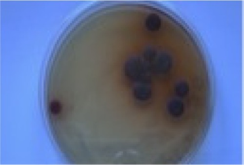
Figure 3 Trichophyton violaceum (white variant).
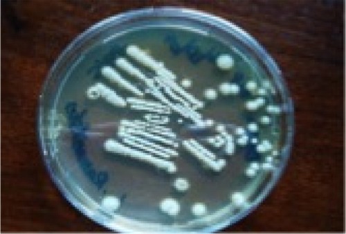
Figure 4 Trichophyton violaceum (lacto phenol cotton blue mount).
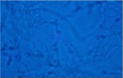
Figure 5 Trichophyton violaceum.
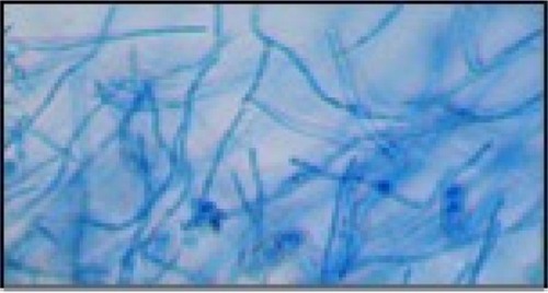
Figure 6 Trichophyton interdigitale.
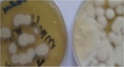
Figure 7 Trichophyton interdigitale (lacto phenol cotton blue mount).
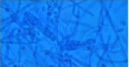
Figure 8 Trichophyton interdigitale.
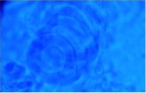
Table 1 Causative dermatophytes in 120 patients
Table 2 Clinical types and causative dermatophytes in 120 patients
Figure 9 Trichophyton tonsurans.
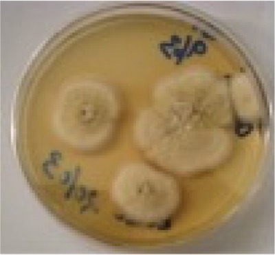
Figure 10 Microsporum ferrugineum (lacto phenol cotton blue mount).
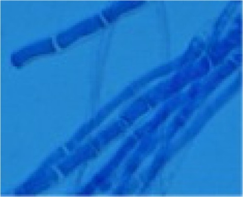
Figure 11 Bar diagram of clinical forms.
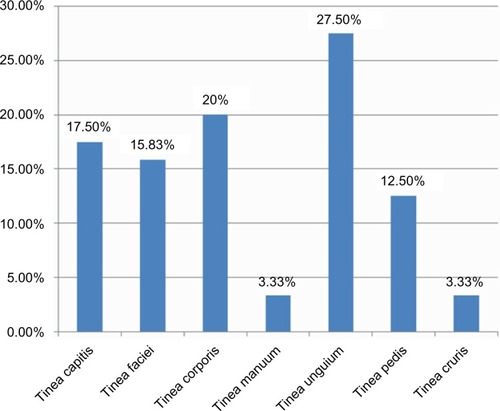
Figure 12 Tinea unguium.
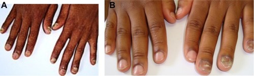
Table 3 Association between age-groups of clinical forms
Figure 13 Tinea corporis (right knee).
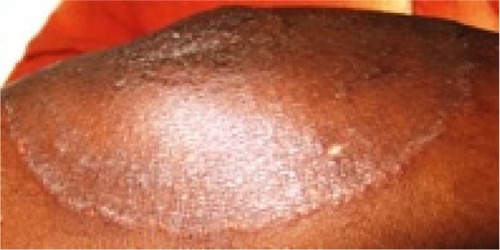
Figure 14 Hair loss, marked crusting and Kerion formation.
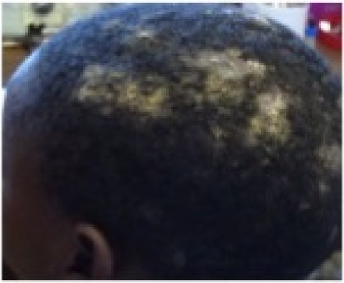
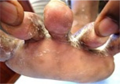
Figure 15 Tinea pedis due to Trichophyton interdigitale.