
Figures & data
Table 1 Histopathological classifications of oral submucous fibrosis
Figure 1 Handmade and commercial forms of betel.
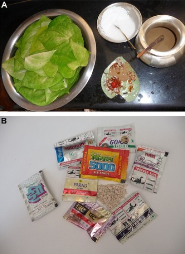
Figure 2 Dental staining and irregular cobble-stone pattern of oral mucosa.
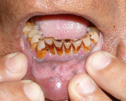
Figure 3 Blanching of buccal mucosa in oral submucous fibrosis.
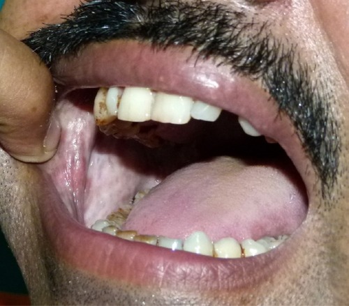
Figure 4 Redness and irregular cobble-stone appearance in oral submucous fibrosis.
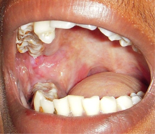
Figure 5 Blanching present on soft palate of patient with oral submucous fibrosis.
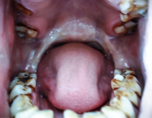
Figure 6 Decreased mouth opening in patient with advanced-stage oral submucous fibrosis.
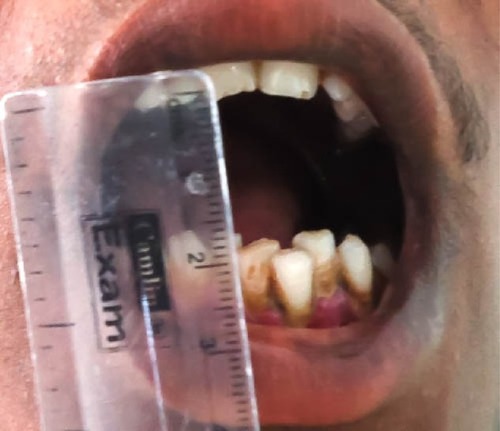
Figure 7 Advanced-stage oral submucous fibrosis.
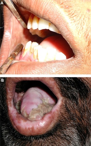
Figure 8 Oral squamous cell carcinoma in a patient with oral submucous fibrosis.
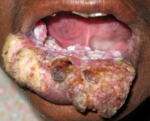
Table 2 Factors associated with malignant transformation of oral submucous fibrosis
Figure 9 Histopathological picture showing initial stage of oral submucous fibrosis.
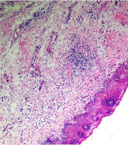
Figure 10 Histopathological picture showing advanced stage of oral submucous fibrosis.
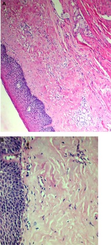
Table 3 Treatment of oral submucous fibrosis (controlled trials)
Table 4 Potential compounds for pharmacological treatment of oral submucous fibrosis