
Figures & data
Figure 1 Techniques for subcutaneous and submucosal PMMA injection.
Abbreviation: PMMA, polymethylmethacrylate.
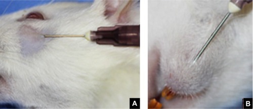
Table 1 Grading scale for inflammatory response
Table 2 Clinical evaluation of local inflammatory response according to PMMA injection site (n=22)
Figure 2 Microscopic appearance after 3 months: few inflammatory cells and formation of connective tissue septa within PMMA spheres.
Abbreviation: PMMA, polymethylmethacrylate.
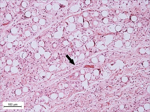
Figure 3 Microscopic appearance 3 months after PMMA injection in the submucosal group.
Abbreviation: PMMA, polymethylmethacrylate.
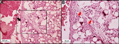
Figure 4 Microscopic appearance of PMMA injection in submucosal group 3 months after PMMA injection, (hematoxylin-eosin, in minor magnification).
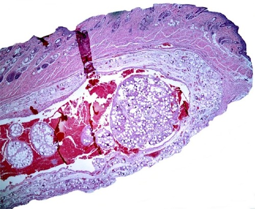
Table 3 Histopathologic evaluation of local inflammatory response according to PMMA injection site
Table 4 Comparative analysis of local inflammatory response to PMMA according to injection site