Figures & data
Table 1 Physical characteristics of liposomes: z-average mean size (d), polydispersity index (PI), zeta potential (ζ), and encapsulation efficiency (E%) for sodium ascorbate
Figure 1 Human skin penetration of empty phosphatidylcholine liposomes.
Notes: Human whole abdomen skin preparations (n=4 per experimental condition) were mounted in Franz cells and exposed to empty phosphatidylcholine liposomes during 0 hour (A), 48 hours (B), and 72 hours (C). Tissue samples were fixed and stained with oil red. Representative microscope images are shown (×400).
Abbreviation: h, hours.
Abbreviation: h, hours.
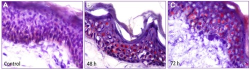
Figure 2 Fluorescein liposome penetration in whole skin.
Notes: (A) Fluorescence microscopy from whole human abdomen skin histology slices after Franz cells time incubation with 1 mg/mL fluorescein liposomes and 70 hours with 1 mg/mL fluorescein solution (image amplification ×200). (B)Time cumulative fluorescein amount in receptor medium of Franz cells after application of fluorescein liposomes (red) or fluorescein solution (yellow). Experiments were performed in skin from four patients and run in triplicate. Results are expressed as mean ± standard deviation.
Abbreviations: min, minutes; h, hours.
Abbreviations: min, minutes; h, hours.
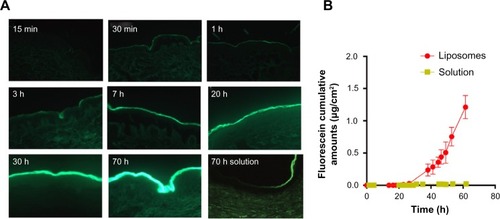
Figure 3 Comparative skin penetration of fluorescein and ascorbate liposomes in different human skin layers.
Notes: (A) Fluorescein and (B) ascorbate liposomes penetration kinetics in epidermis (circle), dermis (square), and whole skin (triangle) of human abdomen. Experiments were conducted in skin from four patients and run in triplicate. Results are expressed as mean ± standard deviation.
Abbreviation: h, hours.
Abbreviation: h, hours.
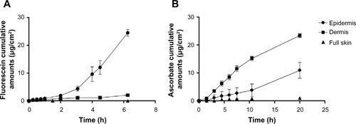
Table 2 Permeability coefficients (Kp) for all formulation tested
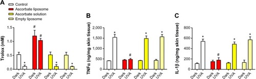
Figure 4 Antioxidant and anti-inflammatory capacity of ascorbate liposomes in human skin irradiated by ultraviolet (UV)A/UVB.
Notes: Human whole abdomen skin was exposed to ascorbate solution or ascorbate liposomes (100 mg/mL) during 20 hours. After washing, skin surface was irradiated with UVA/UVB intensity of 50 J/cm2 for 2 hours and remained incubated in Franz cells for 24 hours. Control preparations were left in Franz cells, in dark conditions. Skin tissue was triturated and homogenized, and antioxidant Trolox capacity (A), tumor necrosis factor alpha (TNFα) (B), and interleukin (IL)-1β (C) were measured by antioxidant and ELISA kits. Experiments were done in skin from four patients and run in triplicate. Results are expressed as mean ± standard deviation. *P<0.05 vs control dark conditions; #P<0.05 vs control group.