Figures & data
Table 1 Summary of the different parameters related to color assessment
Table 2 Color parameters recorded (L*, a*, b*) or calculated (chroma C*, hue angle, and ITA°) from a Chroma-Meter applied on the face skin of 20 volunteers before and after 14, 28, 56, and 84 days of vehicle cream or THBG cream application
Table 3 Melanin content quantification by SIAscope™ applied on the face skin of 20 volunteers at the beginning of the study (D0) and after 14, 28, 56, and 84 days of vehicle and THBG treatment
Figure 1 Melanin content visualization at D0 and after 14, 28, 56, and 84 days of placebo and THBG treatments (SIAscope™ tool, selection of two volunteers).
Abbreviations: THBG, 3,4,5-trihydroxybenzoic acid glucoside; n°, number; D, day.
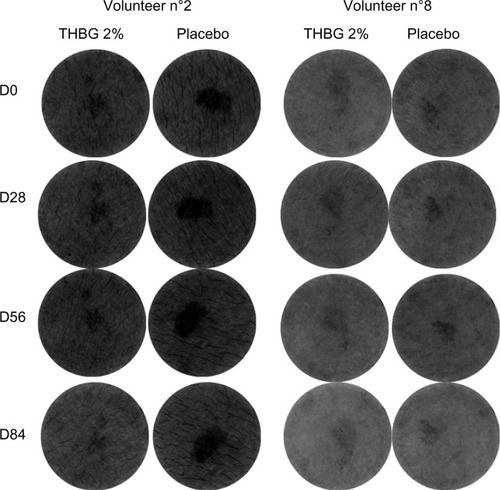
Figure 2 Macrophotographies taken under visible or UV light from one volunteer before and after vehicle cream or THBG cream application for 84 days.
Abbreviations: UV, ultraviolet; THBG, 3,4,5-trihydroxybenzoic acid glucoside; D, day.
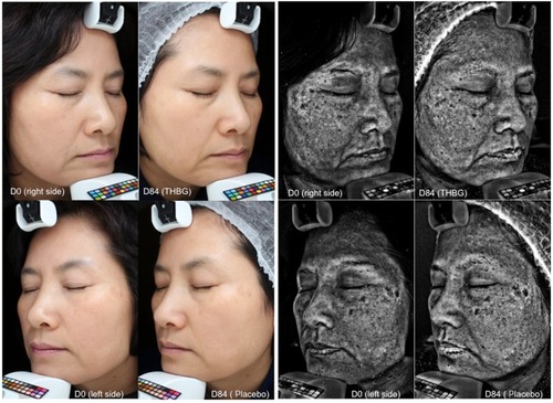
Table 4 UV spots parameters quantification
Figure 3 Macrophotographies taken using visible light from three volunteers before and after vehicle cream or THBG cream application for 84 days.
Abbreviations: THBG, 3,4,5-trihydroxybenzoic acid glucoside; D, day.
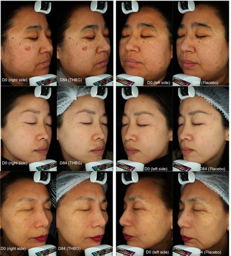
Figure 4 Absorption spectra of THBG pure or diluted in a formulation cream.
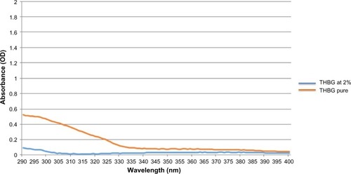
Figure 5 Visualization of melanin content in skin explants cultured in presence or not of THBG at 2% and kojic acid at 1% (histological analysis, Fontana Masson staining).
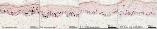
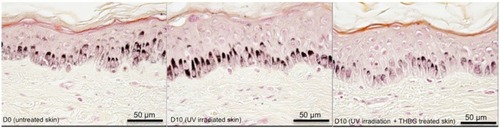
Figure 6 Visualization of melanin content in skin explants cultured after irradiation and in presence or not of THBG at 2% (histological analysis, Fontana Masson staining).