Figures & data
Table 1 Material/Manufacture, Application Mode, Batch Number of Cementation and Adhesive Systems
Table 2 Overall Means and Standard Deviations of the Push-Out Bond Strength Values (MPa) for All Experimental Groups
Table 3 Means and Standard Deviations of the Push-Out Bond Strength Values (MPa) for the Cross-Product Interaction Mode of Application vs Etchant Viscosity (*)
Figure 1 Number of specimens (%) according to the fracture pattern modes observed in the different experimental groups.
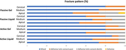
Figure 2 Number of specimens with opened dentinal tubules (%) observed in the different experimental groups. Groups identified with same lowercase letters indicate values statistically similar (p > 0.05).
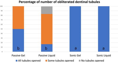
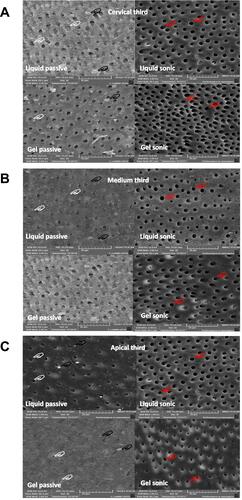
Figure 3 Representative SEM images of each third ((A) cervical; (B) medium; (C) apical) of the conditioning pattern observed in the experimental groups. It was observed that, in all thirds, the liquid and gel applied as passive technique shows dentinal surfaces with smear layer debris (black hands) and partially opened tubules due to the presence of smear plugs (white hands). On the other side, in the groups where the liquid and gel etchants were applied in the sonic mode, it was a more complete opening of the dentinal tubules (red hands).