Figures & data
Table 1 Number and Percentage of Root Canals of Maxillary Second Molars
Table 2 Mean, Standard Deviation, Minimum and Maximum Values of DM, DP in Millimeters and MDP in Degrees in Maxillary Second Molars (n=301)
Table 3 DM (Distance Between the Distobuccal and Mesiobuccal Canal Orifices) Variations in Millimeters
Table 4 DP (Distance Between the Distobuccal and Palatal Canal Orifices) Variations in Millimeters
Table 5 Frequency Distribution of MDP Angle Variations (<90°, 90–120° and >120°)
Table 6 MDP (the Angle Formed by the Mesiobuccal, Distobuccal and Palatal Canal Orifices) Variability
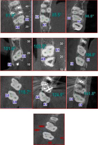
Figure 1 Images of transverse section of the orifice in different cases. From left to right and from top to bottom: MDP<90° and MDP>140°. Blue values show distance between the orifices of distobuccal and mesiobuccal canals (DM), and distance between the orifices of distobuccal and palatal canals (DP). Green values show the angle between the orifices of mesiobuccal, distobuccal and palatal canals (MDP). Red values indicate the canal orifices of four-canal maxillary second molars.