
Figures & data
Figure 1 The schematic representation of the specimen preparation procedures. (A) The crown of a premolar tooth was sectioned and removed. (B) The instrumentation of the root was performed using the ProTaper NiTi rotary system. (C) The final irrigation protocol was performed according to the experimental group. (D) The root was obturated. (E) The roots were embedded in the self-curing acrylic resin block. (F) The fracture resistance was measured.
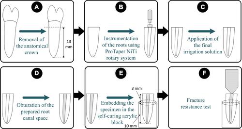
Table 1 The Mean Fracture Resistance Values ± Standard Deviations (N) of the Experimental Groups