
Figures & data
Figure 1 (A) Intraoral view of the upper right quadrant at baseline; calculus and gum bleeding is visible. (B) Intraoral view of the frontal lower teeth at baseline; small amounts of calculus and pus discharge are visible.
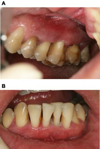
Figure 2 Panoramic radiograph at baseline.
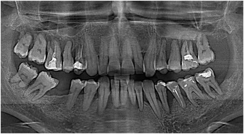
Figure 3 (A) Upper right quadrant after antibiotic therapy. (B) Frontal lower teeth after antibiotic therapy.
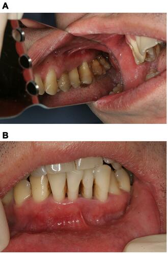
Figure 4 Intraoral view of the frontal lower teeth 14 days after SRP and splinting.
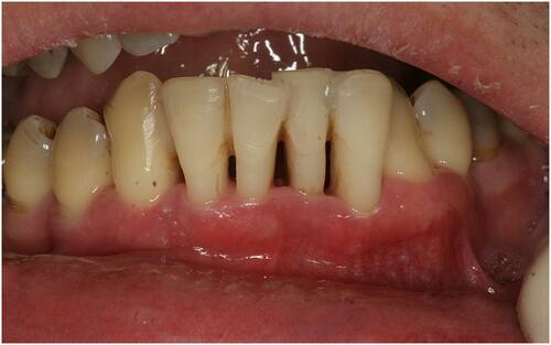
Figure 5 Intraoral view 6 months after SRP.
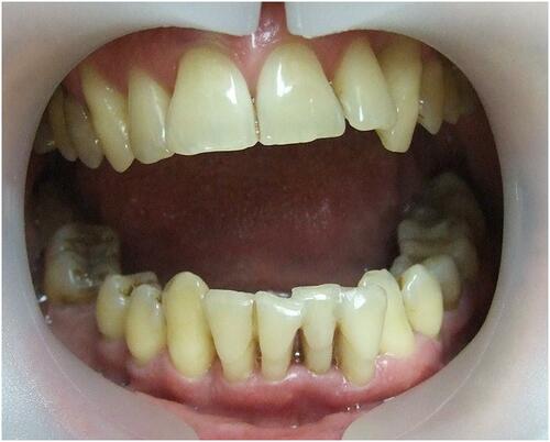
Figure 6 Panoramic radiograph 6 months after SRP: tooth #43 showed an extensive periapical lesion with well-defined borders and bone loss extruding onto the root’s mesial surface.
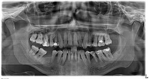
Figure 7 “Walking” probing of the periodontal pocket using a Naber periodontal probe. Distal buccal point (A), middle buccal point (B), mesial buccal point (C), all measurements are 2 mm probing depth. (D) Narrow and deep 8 mm periodontal pocket on the mesial tooth surface.
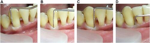
Figure 8 (A) No bleeding was observed immediately after endodontic access opening. (B) Instrumentation of the root canal using an iRace rotary endodontic instrument. (C) Passive ultrasound irrigation with an ultrasound tip. (D) Treating the root canal with ozone gas.
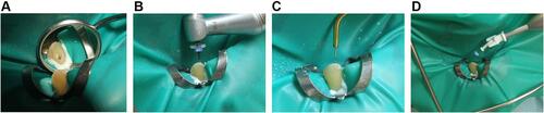
Figure 9 (A) Working length confirmation. (B) Immediately after obturation. (C) Six-month follow-up showing complete healing of the bone defect in the periapical area and interdental septum.
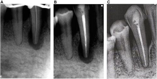
Figure 10 Prozone ozone generator.
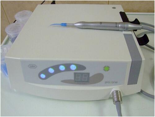
Table 1 Summarizing of Clinical Parameters, Treatment Protocols, and Healing Data of Case Studies with PESP Lesion Type
Table 2 Summarizing of Clinical Parameters, Treatment Protocols, and Healing Data of Case Studies with PPSE Lesion Type
Table 3 Summarizing of Clinical Parameters, Treatment Protocols and Healing Data of Case Studies with Primary Endodontic Diseases
Table 4 Summarizing of Clinical Parameter, Treatment, and Healing Data of a Case with True Combined Lesion