
Figures & data
Table 1 Previous Studies on Evaluation of the Prevalence of MB2 Canals in Different Populations
Figure 1 Diagrammatic representations of Vertucci’s classification for root canal morphology.
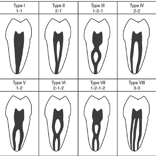
Table 2 Age and Sex Distribution of the Sample
Table 3 Types of Canals on Right and Left Sides
Figure 2 Graphical representation of canal types on the right and left sides.
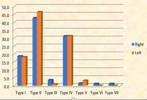
Figure 3 Radiographic representation of canal types I, II, IV, and V in axial and coronal sections on CBCT images (arrows representing position of mesiobuccal canals in MFMs) (Axial 1: Coronal part of the mesiobuccal root in axial section. Axial 2: Middle part of the mesiobuccal root in axial section. Axial 3: Apical part of the mesiobuccal root in axial section). Coronal: coronal section. (A) Type I in axial and coronal sections. (B) Type II in axial and coronal sections. (C) Type IV in axial and coronal sections. (D) Type V in axial and coronal sections.
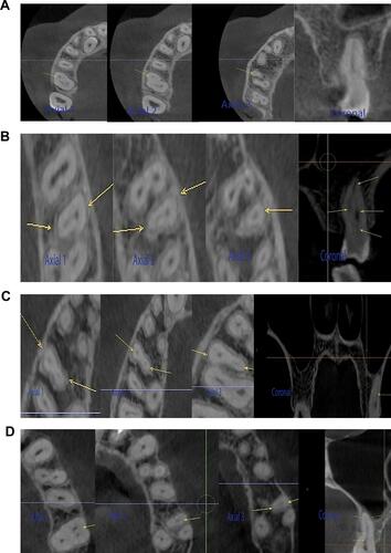
Table 4 Distribution of Canal Type According to Gender
Table 5 Distribution of Types of Canal on the RIGHT Side According to the Age Groups
Table 6 Distribution of Types of Canal on the LEFT Side According to the Age Groups