Figures & data
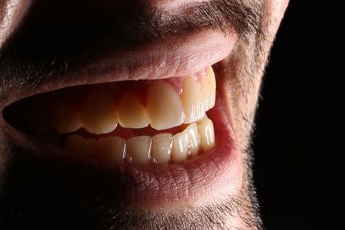
Figure 1 Frontal view of anterior teeth.
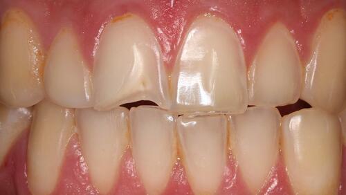
Figure 2 Occlusal view of the maxillary arch.
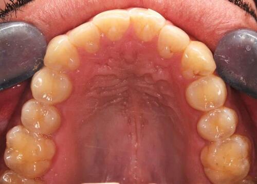
Figure 3 Occlusal view of the mandibular arch.
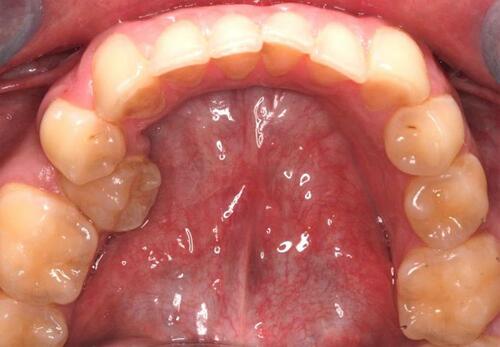
Figure 4 Diagnostic wax-up.
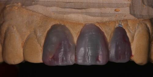
Figure 5 Intraoral mock-up.
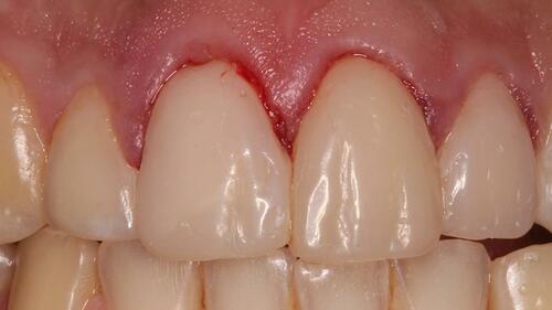
Figure 6 Splint injected with clear PVS and seated on the cast.
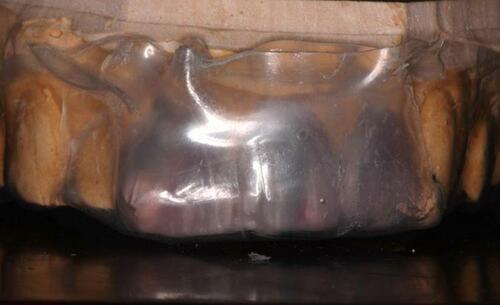
Figure 7 Isolation of adjacent teeth using Teflon tape during restoration of maxillary right central incisor and left lateral incisor.
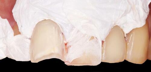
Figure 8 Isolation of adjacent teeth using Teflon tape during restoration of maxillary left central incisor.
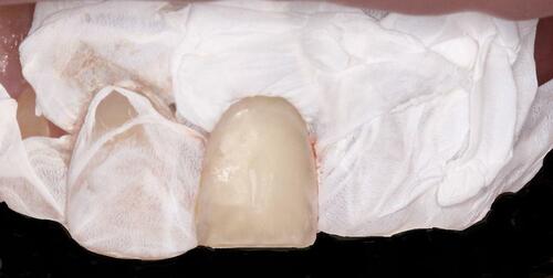
Figure 9 Tip of the injectable composite resin syringe inserted into the space created by the wax-up.
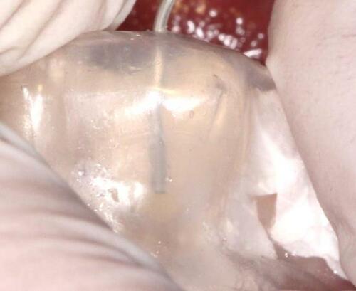
Figure 10 Final restorations.
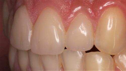
Figure 11 Preoperative smile.
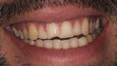
Figure 12 Postoperative smile.
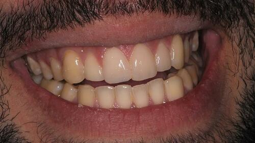
Figure 13 Follow-up photograph after 8 weeks.
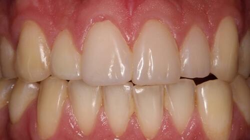
Figure 14 Artistic picture showing the patient’s smile.