Figures & data
Figure 1 Comparative visualization of (A) non-polarized and (B) polarized images. Image (B) highlights the absence of flash reflection on the polarized side.
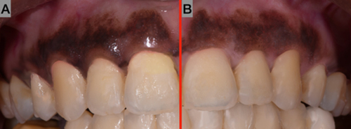
Figure 2 Standardized photos were taken with a stent and a marker to ensure the same position and angle when the images were overlayed over each other.
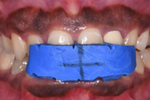
Figure 3 Picture of the full upper arch after gingival depigmentation.
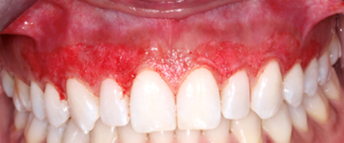
Figure 4 The grid feature in Photoshop 2020 was used to divide each image into numerous smaller samples.
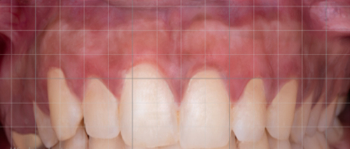
Figure 5 Measurement of gingival pigmentation with the Dummett Oral Pigmentation Index (DOPI) and Gingival Melanosis Record (GMR).
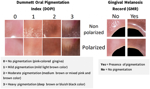
Figure 6 Percentage of GMR of polarized and non-polarized photos among different groups at pre- and post-depigmentation.
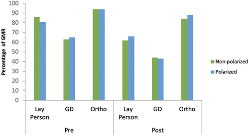
Figure 7 DOPI mean of polarized and non-polarized photos among different evaluators at pre-depigmentation and post-gingival depigmentation.
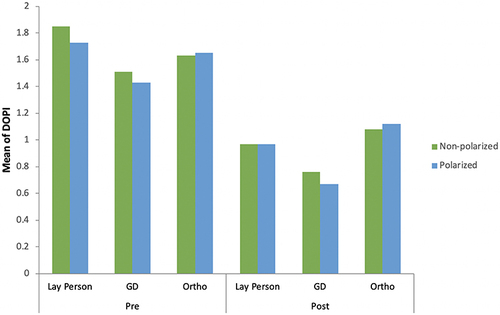
Table 1 Comparison of GMR and DOPI Before and After Depigmentation by Different Evaluators
Figure 8 Percentage of GMR reduction of polarized and non-polarized photos among different groups.
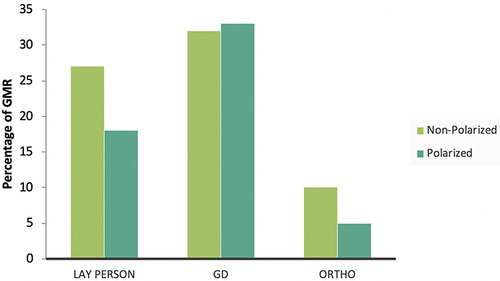
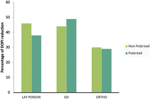
Figure 9 Percentage of DOPI reduction of polarized and non-polarized photos among different groups.