
Figures & data
Figure 1 Transmission electron microscopy image of silver nanoparticles.
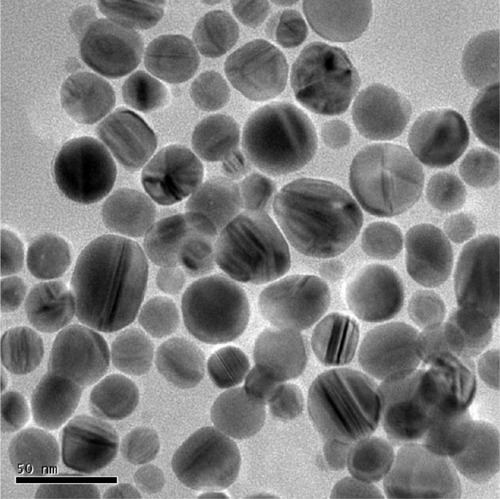
Figure 2 Illustration of the assembly.
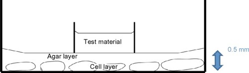
Figure 3 The cytotoxic zone.
Abbreviation: PDL, periodontal ligament.
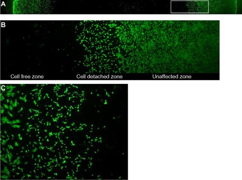
Figure 4 After reducing the diaphragm size of the light source of the phase contrast microscope, at 10× magnification, the field of view covered around 15 PDL cells of about 200 μm diameter.
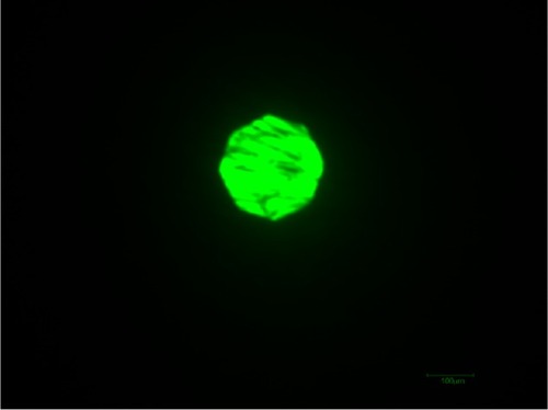
Figure 5 The traveling stage of the microscope was traveling along the diameter of the culture well.
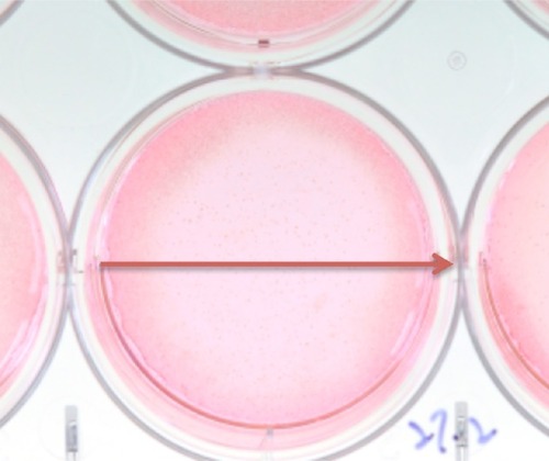
Table 1 Classification of cytotoxic score
Table 2 Interpretation of cytotoxic score
Figure 6 Dose-response relationship between the novel nano-silver irrigant to both NIH 3T3 and hPDLSCs.
Abbreviations: hPDLSCs, human periodontal ligament stem cells; vs, versus.
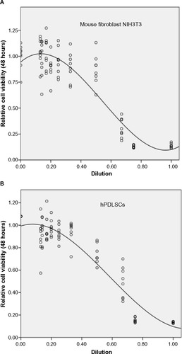
Figure 7 A significant linear relationship of survivability of hPDLSCs and Mouse fibroblast NIH3T3 in different dilutions of nano-silver solution.

Figure 8 Survival curves (generated by the regression model) of hPDLSCs and Mouse fibroblast NIH3T3 (directly cultured for 48 hours with different dilutions of nano-silver).
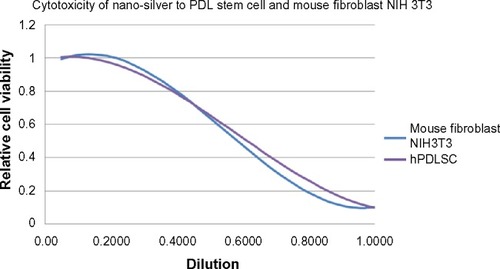
Table 3 Comparison of cytotoxic score of various irrigants on hPDLSCs and Mouse fibroblast NIH3T3