Figures & data
Figure 1 Upper endoscopy demonstrated an ulcerated lesion with an overlying blood clot in the pre-pyloric antrum (arrow).
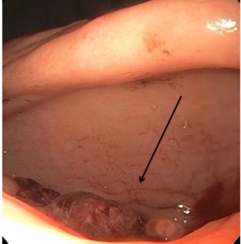
Figure 2 Initial computed tomography of the abdomen revealed significant inflammatory changes around the gallbladder with a large 5 cm gallstone exerting significant mass effect on the antrum of the stomach.
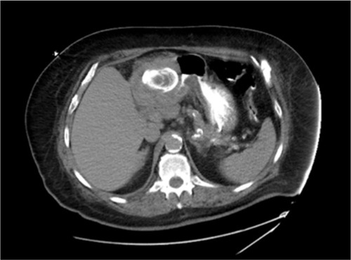
Figure 3 Initial scout films demonstrated a large 5–6 cm gallstone projecting over the left upper quadrant (white arrow; A). Upper gastrointestinal series demonstrated contrast extravasation (black arrow) into the right upper quadrant suspected to be into a contracted gallbladder and consistent with a cholecystogastric fistula (B).
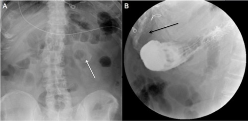
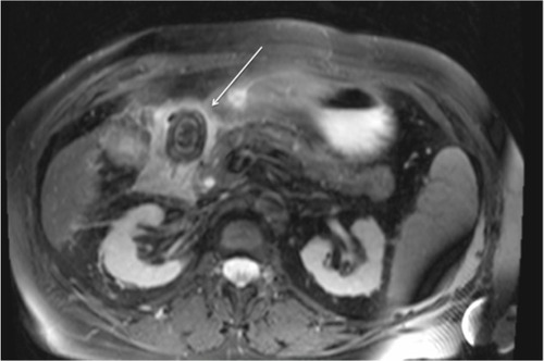
Figure 4 MRI of the abdomen demonstrating a large impacted duodenal gallstone (arrow).