Figures & data
Table 1 Clinicopathological features of patients with rectal varices
Figure 1 A) Cb, F2, RC-positive rectal varices. B) Fluoroscopic observation with infusion of 5% EOI was performed to determine the extent of the varices. C) One week after, fluoroscopic observation with infusion of 5% EOI.
Abbreviations: Cb, blue; EIS, endoscopic injection sclerotherapy; EOI, ethanolamine oleate with iopamidol; F2, enlarged and tortuous; RC, red color.
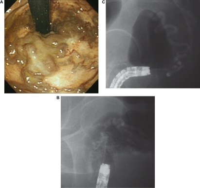
Figure 2 After EIS colonoscopy revealed shrinkage of the rectal varices.
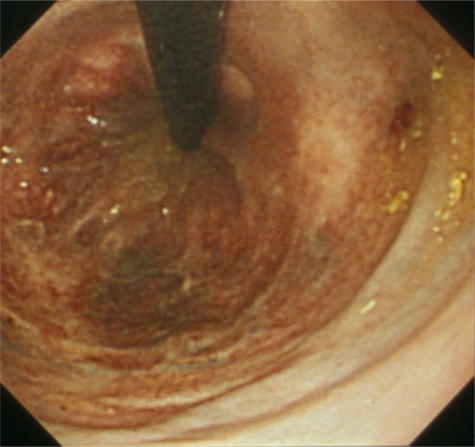
Figure 3 Colonoscopy revealed bleeding from ulcers after endoscopic band ligation.
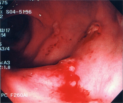
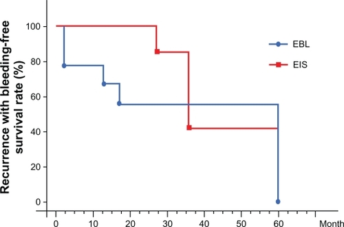
Figure 4 Recurrence with bleeding-free survival rate was calculated by the Kaplan–Meier method for between-group comparisons.