Figures & data
Figure 1 Stewart-Way classification of bile duct injuries.
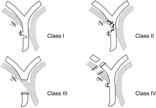
Table 1 Bismuth and Strasberg classification
Table 2 Summary of proposed methods to prevent bile duct injuries
Figure 2 Intra-operative real-time identification of biliary structures with visible light (VL) on left and by fluorescence (NIRF-C) on right.
Abbreviation: NIRF-C, near-infrared fluorescent cholangiography.
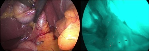
Figure 3 (A) Axial three-dimensional MRCP sequence showing a small fluid collection in the gallbladder fossa (white arrow). (B) T1-weighted hepatobiliary acquisition (after gadoxetic acid injection) demonstrates opacification of the small fluid collection suggesting a biliary leak from the cystic duct.
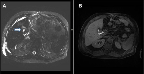
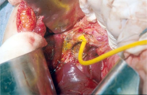
Figure 4 Trans-Kehr cholangiography after the positioning of T-tube to protect biliary anastomotic suture.
Abbreviation: CBD, common bile duct.
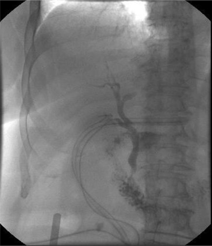
Figure 5 A hepaticojejunostomy was performed for a stricture at biliary confluence after the positioning of T-tube.